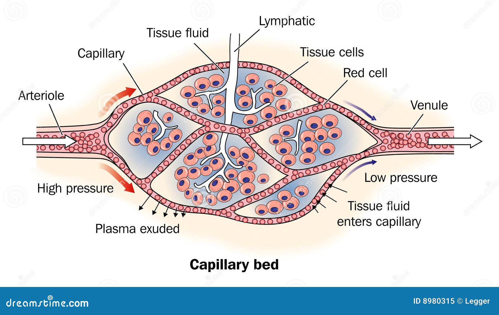
The capillary filtration coefficient dictates how efficiently fluid crosses capillary walls. It measures the volume of fluid filtered across a capillary membrane per unit of time, per unit of pressure gradient. When you study microvascular fluid dynamics, you quickly realize that simple pressure differences don't tell the whole story. Two capillary beds with identical pressure gradients can experience drastically different fluid shifts because their membrane properties vary.
Understanding this coefficient requires looking closely at the Starling equation. Ernest Starling first mapped out how hydrostatic and oncotic pressures govern fluid movement between capillaries and interstitial spaces. Over a century later, modern physiologists continue to refine our understanding of how endothelial glycocalyx layers and surface area constants determine fluid balance in human tissues.
Defining the Starling Equation Foundation
To grasp the mechanics of the capillary filtration coefficient, you have to look at the mathematical framework governing microvascular fluid flux. The net rate of fluid filtration across a capillary wall depends on hydrostatic and osmotic forces combined with membrane permeability characteristics.
Expressed mathematically, fluid movement is calculated through the classical Starling equation:
$$J_v = K_f \cdot [(P_c - P_i) - \sigma(\pi_c - \pi_i)]$$
In this equation, $J_v$ represents the net fluid movement. The term $K_f$ is the coefficient itself. The remaining variables measure capillary hydrostatic pressure ($P_c$), interstitial hydrostatic pressure ($P_i$), the reflection coefficient ($\sigma$), capillary oncotic pressure ($\pi_c$), and interstitial oncotic pressure ($\pi_i$).
Without the $K_f$ term, the equation remains incomplete. It serves as the proportionality constant that bridges the gap between pressure gradients and actual physical volume movement. If $K_f$ is high, even a modest pressure imbalance creates massive fluid shifts. If $K_f$ is low, the capillary wall resists fluid movement despite steep pressure differences.
Key Physiological Determinants of $K_f$
The value of the capillary filtration coefficient isn't static across the human body. Different vascular beds exhibit vastly different baseline permeabilities and surface areas. Two main physiological factors drive these variations.
Capillary Surface Area
Total available surface area dictates how much room fluid has to cross the barrier. Beds with dense capillary networks—such as skeletal muscles during heavy exertion, active renal glomeruli, and pulmonary capillaries—possess exceptionally high surface areas. When precapillary sphincters relax, dormant capillaries open up, which immediately increases the active surface area and raises $K_f$ locally.
Hydraulic Conductivity
Hydraulic conductivity reflects the intrinsic permeability of the capillary wall itself. Continuous capillaries found in skin and muscle feature tight junctions and have low conductivity. Fenestrated capillaries found in the intestines and endocrine glands feature small pores that let water and small solutes pass much more easily. Discontinuous capillaries found in the liver and bone marrow feature wide gaps, resulting in very high hydraulic conductivity.
The Role of the Endothelial Glycocalyx
For decades, classic physiology textbooks taught that fluid reabsorption occurred at the venous end of every capillary because interstitial oncotic pressure pulled fluid back in. Modern microvascular research has upended that view, largely due to our understanding of the endothelial glycocalyx.
The glycocalyx is a gel-like carbohydrate-protein layer lining the luminal surface of endothelial cells. It acts as a primary filter. Under normal conditions, the subglycocalyx space has a very low oncotic pressure. This means fluid reabsorption across the traditional capillary wall is much lower than older models predicted.
When inflammatory mediators degrade the glycocalyx, the physical barrier breaks down. The effective filtration surface changes, and $K_f$ spikes. This loss of barrier integrity explains why systemic inflammation leads to rapid edema formation in tissues like the lungs and skin.
Clinical Implications and Pathological Shifts
Changes in the capillary filtration coefficient sit at the center of several acute and chronic medical conditions. When the microvascular barrier fails, clinical management becomes a race to control fluid shifts.
Pulmonary Edema and ARDS
In acute respiratory distress syndrome, inflammatory cytokines damage the alveolar-capillary membrane. This injury increases the hydraulic conductivity component of $K_f$. As a result, normal pulmonary capillary pressures force massive amounts of fluid out of the bloodstream and into the air spaces of the lungs. Gas exchange collapses because the diffusion distance for oxygen widens dramatically.
Chronic Heart Failure
In chronic congestive heart failure, long-term venous hypertension elevates capillary hydrostatic pressure ($P_c$). Over time, the microvasculature adapts. Chronic high pressure can alter the structural integrity of the capillary wall, modifying local fluid exchange parameters and worsening peripheral edema in the lower extremities.
Burn Injuries
Severe thermal injuries trigger systemic capillary leak syndrome. Plasma proteins escape into the interstitial spaces because $K_f$ increases sharply across large skin surfaces. Fluid resuscitation protocols must account for this sudden spike in microvascular permeability to prevent hypovolemic shock.
Measuring and Estimating Filtration Rates
Quantifying the capillary filtration coefficient in living human tissues presents significant technical hurdles. Researchers typically rely on specialized plethysmography in limbs or isolated organ perfusion models in experimental settings.
By abruptly increasing venous pressure—such as by inflating a cuff on a forearm—investigators can measure the initial rate of fluid filtration into the hand or forearm tissues. Because arterial pressure and oncotic pressures remain momentarily stable, the sudden change in volume reflects the underlying filtration capacity. This technique provides reliable estimates of microvascular compliance and permeability changes in clinical research environments.
You can explore more detailed physiological parameters and clinical research through resources provided by the National Center for Biotechnology Information or academic overviews hosted by PhysioPedia.
Frequently Asked Questions
What units are used to measure the capillary filtration coefficient?
It is typically expressed in milliliters per minute per millimeter of mercury per 100 grams of tissue ($mL \cdot min^{-1} \cdot mmHg^{-1} \cdot 100g^{-1}$). This standardizes measurements across different tissue masses and pressure scales.
Why do some organs have higher filtration coefficients than others?
Organs with specialized filtration jobs—like the kidneys, which must filter massive volumes of plasma to form urine—require high hydraulic conductivity and large surface areas. In contrast, tissues that protect internal environments, such as the blood-brain barrier, maintain extremely low coefficients to restrict unwanted fluid and solute movement.
Does the capillary filtration coefficient change during exercise?
Yes. During vigorous exercise, metabolic vasodilation recruits dormant capillaries in skeletal muscle. This recruitment expands the total surface area available for fluid and nutrient exchange, raising the effective $K_f$ in those muscle beds.
How does inflammation affect microvascular fluid exchange?
Inflammation releases histamine, bradykinin, and cytokines that degrade the endothelial glycocalyx and widen gaps between endothelial cells. This structural damage increases hydraulic conductivity, driving fluid out of the capillaries and into the interstitial space to cause localized swelling.

