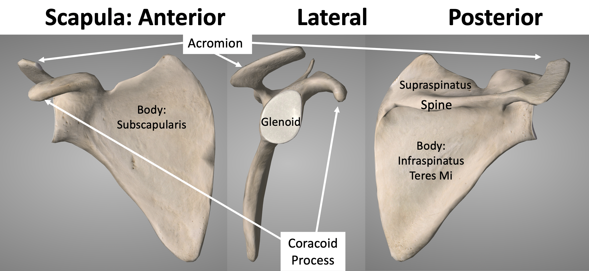
The acromion is the bony extension of the scapula forming the roof of your shoulder joint. It's the structural anchor where your collarbone meets the shoulder blade, creating a protective vault for the rotator cuff tendons underneath. When this architecture narrows or irritates the soft tissues below, it triggers chronic pain and limits upper body mobility. Understanding how this bone works helps explain why common shoulder injuries happen and how clinicians treat them.
Anatomically, the acromion projects laterally and superiorly off the spine of the scapula. It articulates directly with the lateral end of the clavicle to form the acromioclavicular joint, a crucial connection that stabilizes the entire shoulder girdle during lifting and reaching movements. The shape of this bone varies significantly from person to person, falling into three distinct morphological categories that directly influence physical health and injury vulnerability.
Acromion Types and Structural Variations
Orthopedic specialists classify acromial morphology into three distinct shapes based on imaging studies. These structural variations determine how much clearance exists between the bone and the soft tissues traveling through the subacromial space.
- Type I acromions feature a flat underside, offering ample clearance for the rotator cuff tendons and the subacromial bursa to glide smoothly during arm elevation.
- Type II acromions present a curved profile that mirrors the rounded contour of the humeral head, moderately reducing the working space beneath the bone.
- Type III acromions exhibit a downward-projecting hook at the anterior tip, creating a mechanical bottleneck that frequently abrades the supraspinatus tendon.
People born with a type III acromion face a much higher statistical likelihood of developing chronic mechanical wear. Over decades of repetitive overhead motion, this hooked tip acts like a sandpaper edge against the underlying tendon fibers. Structural shape doesn't change on its own, but bone spurs can develop over time on any acromion type due to chronic traction from the coracoacromial ligament, further narrowing the available clearance.
Biomechanics and Scapulohumeral Rhythm
Your shoulder relies on intricate kinetic coordination known as scapulohumeral rhythm. As you raise your arm overhead, the humerus moves in tandem with the scapula, and the acromion rotates upward to keep from pinching the rotator cuff.
When you lift your arm past ninety degrees of abduction, the greater tuberosity of the humerus comes within millimeters of the acromion's undersurface. Without proper upward rotation of the scapula driven by the serratus anterior and trapezius muscles, that clearance vanishes instantly. This pinching action happens thousands of times a day in professions requiring overhead work, swimming, or weightlifting, placing immense cyclic fatigue on the supraspinatus tendon.
The deltoid muscle also originates directly from the anterior and lateral borders of the acromion. When the deltoid contracts to elevate the arm, it pulls upward on the humerus while simultaneously creating an upward shear force against the acromion itself. The rotator cuff muscles must actively depress the humeral head downward to counteract this upward pull, maintaining joint centering. If the rotator cuff fatigues, the humeral head migrates superiorly, crashing directly into the acromion.
Clinical Pathologies Linked to the Acromion
Structural friction and trauma around the acromion give rise to several well-documented orthopedic conditions that disrupt daily upper extremity function.
Subacromial Impingement Syndrome
This condition occurs when repetitive mechanical compression irritates the structures housed in the subacromial space, primarily the supraspinatus tendon and the subacromial bursa. Patients typically report a painful arc of motion when raising the arm between sixty and one hundred twenty degrees. Over time, persistent inflammation leads to tendinosis, partial-thickness tears, and eventually full-thickness rotator cuff ruptures if left unaddressed. You can read more about clinical diagnostic criteria through the American Academy of Orthopaedic Surgeons.
Acromioclavicular Joint Arthritis
Because the acromion forms one side of the acromioclavicular joint, degenerative changes frequently target this junction. Osteoarthritis here stems from chronic loading, previous separation injuries, or normal aging. Cartilage wears away, osteophytes form along the joint margins, and local inflammation causes sharp pain at the very top of the shoulder, especially when cross-body adduction forces the joint surfaces together.
Acromion Fractures
Trauma to the lateral shoulder, such as a direct blow from a fall or a high-impact collision, can fracture the acromion process. These fractures compromise the stability of the shoulder girdle and often disrupt the coracoclavicular ligaments. Depending on the displacement pattern, clinicians may manage stable cracks with a simple sling, while displaced fragments often require surgical fixation to restore proper deltoid tension and contour.
Diagnostic Imaging and Evaluation
Pinpointing structural issues involving the acromion requires a combination of physical examination and advanced imaging modalities. Standard radiographs provide the initial baseline. True scapular Y-views and outlet views let physicians visualize acromial slope and spot subacromial bone spurs or joint space narrowing.
When soft tissue damage or subtle stress fractures are suspected, magnetic resonance imaging offers superior visualization of the rotator cuff tendons and the subacromial bursa. According to guidelines from the National Institutes of Health, detailed imaging helps differentiate between acute inflammatory bursitis and degenerative tendon tearing, guiding whether conservative rehabilitation or surgical intervention is appropriate.
Modern Management and Rehabilitation
Treatment strategies for acromion-related disorders have shifted away from aggressive surgical bone shaving toward targeted conservative care. Physical therapy focuses on correcting scapular dyskinesis, strengthening the dynamic stabilizers of the rotator cuff, and opening up the subacromial space through postural adjustments.
When surgery is necessary, such as arthroscopic subacromial decompression for refractory cases, rehabilitation emphasizes early passive range of motion followed by progressive rotator cuff strengthening. Patient recovery timelines depend heavily on adhering to structured physical therapy protocols that restore normal scapulohumeral rhythm without overloading healing tissues.
Frequently Asked Questions
Can you change the shape of your acromion through exercise?
No. Exercise cannot alter your skeletal bone morphology or smooth out a hooked acromion type III. However, targeted physical therapy can strengthen the muscles that control scapular rotation, increasing the dynamic clearance between the acromion and the humeral head to prevent impingement pain.
What is the difference between a bone spur and a hooked acromion?
A hooked acromion is a congenital anatomical variation in the curvature of the bone present from skeletal maturity. A bone spur is a calcific deposit that forms later in life along the edge of the bone or ligament attachment due to chronic mechanical stress and inflammation.
Why does shoulder impingement hurt more at night?
Nighttime pain is common because lying flat eliminates the gravitational assistance that pulls the humeral head downward during the day. In bed, the shoulder joint capsule often relaxes or shifts, allowing the humeral head to migrate upward and press against the acromion and inflamed bursa.
How long does recovery take after acromion decompression surgery?
Basic range of motion usually returns within four to six weeks, but full recovery and return to heavy overhead lifting or sports typically require three to six months of dedicated physical therapy to rebuild strength and dynamic stability.