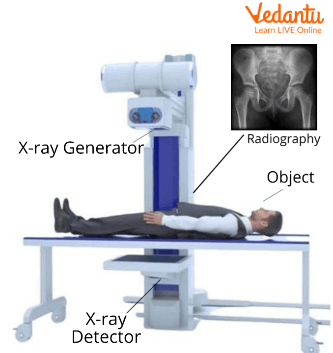
X-ray holes and precision micro-apertures control radiation pathways in advanced imaging systems, allowing technicians to inspect internal structures without destructive testing. Without these controlled optical pathways, high-energy beams would scatter, destroying image resolution in industrial non-destructive testing (NDT) and hospital diagnostics. Understanding how these apertures function helps engineers optimize equipment performance across aerospace, electronics, and clinical sectors.
Modern manufacturing and healthcare rely heavily on clarity. When you inspect multi-layer printed circuit boards, cast turbine blades, or dense skeletal tissue, ray containment dictates success. This article breaks down how precision apertures work, where they are applied, and how engineers maintain them under strict operational standards.
Mechanics of X-Ray Apertures and Focal Paths
Generating clean radiation requires directing electrons through a tightly managed sequence. Inside an industrial or medical X-ray tube, electrons accelerate from a heated filament toward a target anode.
The beam passes through an engineered aperture—frequently referred to as a micro-focus hole or collimation tunnel—to restrict scatter. To understand the complete picture, check out the excellent analysis by The New York Times.
- Electron Focusing: Magnetic lenses concentrate the electron stream down to a diameter of a few micrometers before it strikes the tungsten target.
- Beam Collimation: Physical lead or tungsten apertures shape the resulting radiation cone, preventing peripheral rays from blurring the digital detector array.
- Thermal Dissipation: Because concentrated energy generates extreme heat, aperture structures rely on high-conductivity backing plates, such as diamond or copper alloys, to maintain structural stability.
This precise routing ensures that the shadow projection captured by digital detectors retains high contrast resolution. If the aperture wears down or drifts out of alignment, geometric unsharpness ruins the scan.
Industrial Inspection Applications
Non-destructive testing uses micro-aperture imaging to uncover internal defects before components fail in the field. Industries with zero-failure tolerances rely on these systems daily.
Electronics and Semiconductor Manufacturing
Printed circuit boards feature hidden solder joints underneath ball grid array (BGA) packages. Optical cameras cannot see beneath these chips. X-ray inspection systems use fine focal spots to peer through packaging, exposing micro-voids, cold solder joints, and internal bridging.
Aerospace and Automotive Castings
Critical components like turbine blades and aluminum engine blocks undergo rigorous radiographic screening. Internal gas pockets, slag inclusions, or shrinkage cracks weaken metal structures under stress. Controlled beam tunnels allow inspectors to view these volumetric flaws in three dimensions via computed tomography (CT) without ruining expensive parts.
For more details on industrial testing methodologies, consult the Baker Hughes NDT Solutions Guide.
Medical Diagnostics and Imaging Precision
In clinical environments, radiation exposure must remain as low as reasonably achievable (ALARA) while maximizing diagnostic clarity. Aperture design directly influences patient safety and image sharpness.
- Computed Tomography (CT) Scanners: Gantry systems use precision collimation slits to shape the fan beam, reducing scatter radiation from reaching the patient and improving soft-tissue contrast.
- Fluoroscopy: Real-time imaging procedures depend on stable focal spots so surgeons can navigate catheters and orthopedic hardware with millimeter accuracy.
- Mammography: Specialized micro-focus tubes highlight microcalcifications, which serve as early indicators of malignant tissue changes.
Maintaining Aperture Integrity and Compliance
Over time, continuous exposure to high-energy radiation degrades tube components. Focal spot drift and aperture pitting cause image degradation, leading to false positives or missed defects. Modern facilities follow strict protocols to keep inspection systems calibrated.
- Regular Profiling: Technicians use edge-test methods and digital profile analysis to measure focal spot dimensions, ensuring they stay within micro-tolerance thresholds.
- Thermal Monitoring: Systems track internal operating temperatures to prevent micro-cracking in the tungsten target and surrounding aperture shields.
- Standard Adherence: Operators align maintenance schedules with recognized frameworks such as ISO NDT Standards to verify equipment repeatability.
Neglecting these maintenance steps introduces severe operational risks, including sudden equipment failure during production runs and costly regulatory non-compliance.
Frequently Asked Questions
What causes an X-ray focal spot to degrade over time?
Continuous electron bombardment causes thermal stress and gradual erosion of the tungsten target layer. This wear widens the effective focal spot, increasing geometric unsharpness in digital images.
How do micro-focus systems differ from standard X-ray tubes?
Micro-focus systems use electromagnetic lenses and precise physical apertures to shrink the electron beam to micrometers. Standard tubes have broader focal spots, which limit magnification capabilities because edges blur when enlarged.
Can X-ray inspection detect internal cracks in composite materials?
Yes. Modern high-resolution computed tomography utilizes fine beam collimation to detect fiber misalignment, delamination, and tiny voids inside carbon-fiber composites.
Why is digital detector array technology replacing traditional film?
Digital arrays offer superior contrast resolution, eliminate chemical processing delays, and allow technicians to manipulate image contrast instantly for more accurate flaw detection.