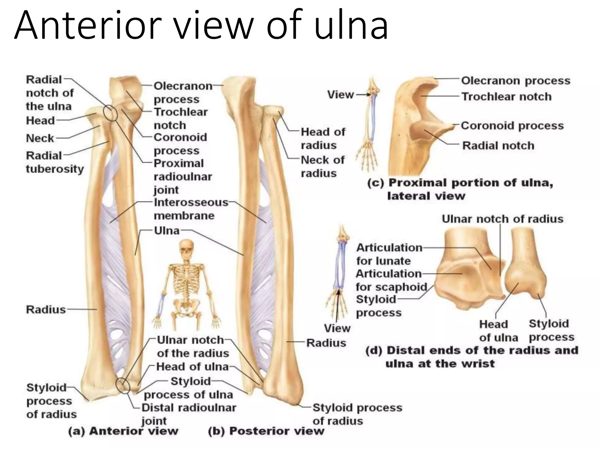
Treating a radius shaft fracture requires precise anatomical restoration to preserve the complex rotational mechanics of the forearm. When surgeons utilize the AO (Arbeitsgemeinschaft für Osteosynthesefragen) approach, the primary goal isn't just bone healing; it's restoring the unique radial bow and maintaining full pronation and supination. Standard casting often fails to maintain this exact alignment, making open reduction and internal fixation (ORIF) the gold standard for displaced adult fractures.
Choosing the correct surgical window dictates success. Surgeons typically rely on either the anterior Henry approach or the posterolateral Thompson approach, depending on the exact location of the break along the radial shaft. Mastering these tissue intervals protects critical neurovascular structures while allowing direct access to the bone for stable compression plating.
Surgical Approaches and Anatomical Landmarks
Gaining exposure to the radius demands a thorough understanding of forearm fascial planes. Entering through the wrong interval risks damaging the posterior interosseous nerve or the radial artery.
The anterior (Henry) approach provides versatile access to the entire length of the radius. Proximal landmarks include the biceps tendon medial to the brachioradialis, while the distal landmark is the radial styloid process. Surgeons develop the interval between the brachioradialis and the flexor carpi radialis. Deep dissection requires identifying the radial artery and its venae comitantes, ligating lateral recurrent branches, and retracting the superficial radial nerve safely.
For middle and distal third fractures, the posterolateral (Thompson) approach offers an alternative route. This method utilizes the interval between the extensor carpi radialis brevis and the extensor digitorum communis. Surgeons must carefully locate and protect the posterior interosseous nerve as it pierces the supinator muscle. Full supination during proximal exposure safely displaces this nerve away from the surgical field.
According to the AO Foundation Surgery Reference, preserving soft-tissue attachments during these exposures minimizes devascularization and promotes primary bone healing.
AO Principles of Internal Fixation
The AO foundation revolutionized fracture management by establishing four core principles: anatomical reduction, stable fixation, preservation of blood supply, and early functional mobilization. For radius shaft fractures, achieving anatomical reduction means restoring length, axis, and rotation precisely.
When both bones of the forearm are broken, surgical sequencing matters. Most trauma protocols dictate that surgeons address the ulna first to establish baseline length, followed by definitive fixation of the radius. However, if the radius fracture dictates overall length and comminution is minimal, stabilizing the radius first can re-establish the structural template.
Compression plating remains the workhorse technique for transverse and short oblique fractures. Applying a 3.5 mm dynamic compression plate (DCP) or limited-contact dynamic compression plate (LC-DCP) generates absolute stability across the fracture site. Lag screws can be added independently when fracture obliquity exceeds twice the width of the bone shaft, enhancing interfragmentary compression before plate application.
Managing Complicated Patterns and Comminution
Not all fractures fit simple transverse patterns. Highly comminuted or segmental radius shaft fractures challenge the surgeon's ability to achieve absolute stability through direct compression alone.
In cases with significant bone loss, bridge plating techniques preserve biological healing by bypassing the zone of comminution entirely. This maintains overall radial length and alignment without stripping soft tissue from small, intermediate bone fragments. Autogenous cancellous bone grafting is often integrated into these procedures to stimulate osteogenesis when cortical gaps threaten delayed union or nonunion.
Surgeons also monitor closely for synostosis—aberrant bone bridging between the radius and ulna. Utilizing separate incisions for the radius and ulna significantly reduces this complication compared to single-incision techniques.
For a deeper look into clinical classification and treatment algorithms, consult Orthobullets Trauma Overview.
Postoperative Rehabilitation and Functional Recovery
Rigid internal fixation permits early controlled motion, which prevents joint stiffness and tendon adhesions. Heavy lifting and resisted exercises remain strictly prohibited until bridging callus appears on follow-up radiographs, usually around six to twelve weeks post-surgery.
Physical therapy focuses on restoring terminal pronation and supination early in the recovery window. Supervised active-assisted range of motion exercises begin as soon as soft tissues tolerate initial swelling reduction, typically within the first two weeks.
Frequently Asked Questions
Why is restoring the radial bow critical during surgery?
The radial bow is a gentle curve essential for the forearm to rotate smoothly over the ulna. If a plate flattens this curve during fixation, the radius binds against the ulna, permanently restricting pronation and supination.
When is bone grafting necessary for a radius shaft fracture?
Bone grafting is indicated when there is segmental bone loss, significant comminution, or atrophic nonunion. Autologous cancellous bone harvested from the iliac crest provides osteoinductive properties that accelerate cortical healing.
What is the risk of a single-incision approach for both forearm bones?
Using one incision to plate both the radius and ulna dramatically increases the incidence of radioulnar synostosis, where bone bridges the two shafts and locks forearm rotation. Two separate incisions mitigate this risk.
How long do plates remain in place after fixation?
Implants are generally left in place unless they cause localized irritation, particularly along the subcutaneous border of the ulna. If removal is planned, it shouldn't occur earlier than two years post-osteosynthesis to ensure complete cortical remodeling.