The phosphate backbone forms the continuous structural spine of the DNA double helix. Without it, the genetic instructions required for cellular replication, protein synthesis, and organismal inheritance would lack physical stability. Far from being a passive structural rope, this alternating chain of sugar molecules and negatively charged phosphate groups actively dictates how nucleic acids interact with water, ions, and nuclear enzymes.
Understanding the atomic architecture of this backbone clarifies how DNA resists physical shearing while remaining flexible enough to unwind during transcription. By examining phosphodiester bonds, charge distribution, and conformational adaptability, we can see how nature engineers molecular stability at the nanoscale.
Chemical Architecture: Phosphodiester Bonds and Sugar-Phosphate Polarity
The backbone consists of repeating units of deoxyribose sugar and phosphate groups linked together by covalent phosphodiester bonds. Each phosphate group connects the 3' carbon atom of one sugar molecule to the 5' carbon atom of the next. This directional linkage establishes the inherent polarity of a DNA strand, creating distinct 5' and 3' ends.
The repeating covalent sequence runs along the outside of the double helix, while the nitrogenous bases (adenine, thymine, cytosine, and guanine) stack perpendicular to the central axis on the inside. This spatial arrangement shields the hydrophobic core of base pairs from the surrounding aqueous cellular environment while exposing the hydrophilic phosphate groups to water molecules.
Directionality dictates how cellular machinery reads and copies genetic material. Enzymes such as DNA polymerase can only synthesize new strands in the 5' to 3' direction because of the specific chemical orientation of the free hydroxyl group on the 3' carbon and the triphosphate group on the incoming nucleotide.
Electrostatic Forces and Ionic Shielding
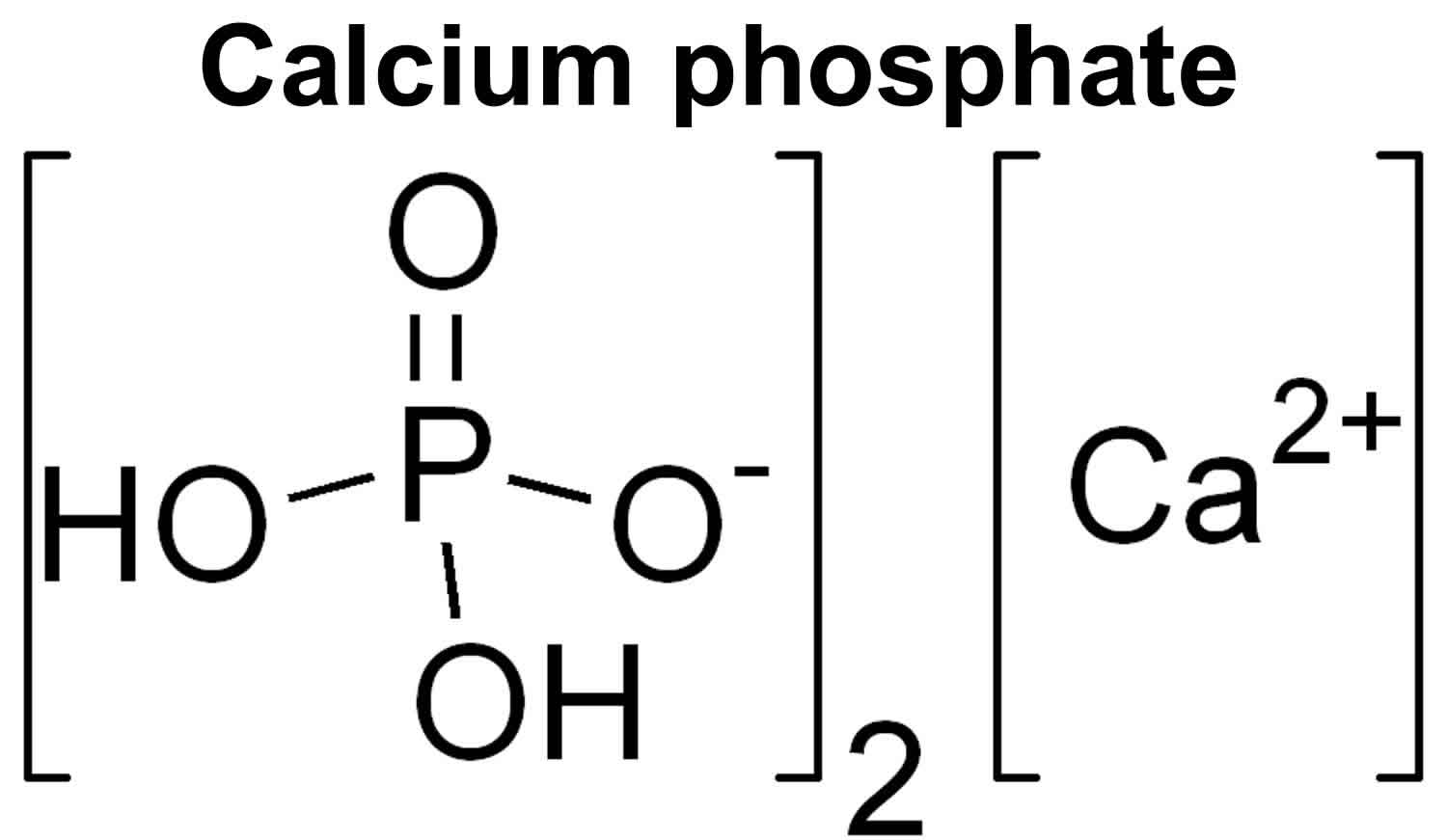
Each phosphate group in the backbone carries a negative charge at physiological pH due to the ionization of its oxygen atoms. This uniform negative charge density creates strong electrostatic repulsion between the two parallel strands of the double helix.
To prevent this repulsion from tearing the molecule apart, cellular fluids contain high concentrations of positively charged counter-ions, primarily magnesium ions, potassium ions, and sodium ions. These cations cluster around the oxygen atoms of the phosphate groups, neutralizing their repulsive forces and allowing the double helix to remain tightly coiled.
Electrostatic tuning also governs how DNA interacts with proteins. Histone proteins, which package DNA into compact chromatin within the nucleus, are rich in positively charged amino acids like lysine and arginine. These basic residues form ionic bonds with the negatively charged oxygen atoms of the phosphate backbone, anchoring the DNA tightly without sequence-specific restrictions.
Conformational Flexibility and Structural Transitions
While the phosphodiester backbone provides rigidity, it possesses enough torsional flexibility to undergo significant conformational shifts. DNA does not exist as a single, rigid rod; it shifts between structural forms depending on hydration levels and protein binding activity.
The B-DNA form represents the standard physiological conformation, featuring a wide major groove and a narrow minor groove that allow transcription factors to read base sequences without breaking the backbone. Under different cellular conditions or in dehydrated states, DNA can transition into A-DNA, which is shorter and wider, or Z-DNA, a left-handed helix characterized by a zig-zag phosphate backbone.
Local backbone flexibility is equally critical during enzymatic processes. When helicase enzymes unzip the double helix during replication, or when restriction enzymes bend DNA to cut specific sequences, the sugar-phosphate backbone twists and distorts without snapping. This resilience is made possible by slight rotations around the single bonds of the sugar-phosphate chain.
Common Misunderstandings About DNA Structure
It's easy to misinterpret diagrams of the double helix and misunderstand how the backbone functions. Here are a few frequent misconceptions:
- The backbone holds the two strands together via covalent bonds: The covalent phosphodiester bonds only hold individual nucleotides together along a single strand. The two complementary strands are held together by much weaker hydrogen bonds between the nitrogenous bases and hydrophobic stacking interactions.
- The backbone is rigid like a metal rod: The sugar-phosphate chain is flexible, capable of localized bending, twisting, and stretching to accommodate protein binding and supercoiling.
- DNA charge is neutral: The backbone imparts a strongly negative net charge to every nucleic acid molecule, which is why techniques like gel electrophoresis successfully pull DNA through a matrix toward a positive electrode.
Applications in Biotechnology and Nanotechnology
Engineers and molecular biologists exploit the predictable properties of the phosphate backbone to design novel biotechnological tools. In CRISPR-Cas9 gene editing, guide RNA molecules rely on exact spatial alignment with target DNA backbones to position Cas9 for precise double-strand cleavage.
In the field of DNA nanotechnology, researchers use synthetic single-stranded DNA sequences—often called scaffold strands—to fold into complex two-dimensional and three-dimensional shapes through programmable base pairing. Because the backbone's charge and flexibility are uniform and predictable, scientists can engineer nanoscale containers for targeted drug delivery, biosensors, and molecular computing circuits.
Furthermore, modifications to the phosphate backbone are foundational in pharmaceutical development. Antisense oligonucleotides and therapeutic mRNA vaccines frequently incorporate modified backbones, such as phosphorothioate linkages where a sulfur atom replaces a non-bridging oxygen. This simple atomic swap protects the synthetic strands from rapid degradation by cellular nucleases, prolonging their therapeutic lifespan in the body.
Frequently Asked Questions
What role does the sugar component play in the backbone?
The deoxyribose sugar acts as the structural connector between the phosphate group and the nitrogenous base. Its 5-carbon ring structure determines the spatial geometry of the backbone and ensures the correct spacing between consecutive base pairs, maintaining the uniform diameter of the double helix.
Why is the phosphate backbone on the outside of the DNA molecule?
Placing the charged phosphate groups on the exterior maximizes their favorable interactions with surrounding water molecules and cellular ions. Meanwhile, the hydrophobic nitrogenous bases are tucked safely into the interior, shielded from water to stabilize the overall molecular structure.
How do mutations affect the phosphate backbone?
Most genetic mutations occur in the nitrogenous base sequence rather than the backbone itself, because the phosphodiester bond pattern is chemically uniform across all genes. However, certain types of DNA damage, such as single-strand or double-strand breaks caused by ionizing radiation, directly sever the backbone, requiring rapid intervention by cellular DNA repair enzymes like DNA ligase.
Can synthetic organisms use a different backbone?
Yes. Synthetic biologists have successfully synthesized xeno-nucleic acids (XNAs), which replace the natural deoxyribose-phosphate backbone with alternative molecular chains, such as threose or cyclohexene rings. These synthetic polymers demonstrate that life's core information storage system can function on modified physical scaffolds.