Proteins drive nearly every cellular process in living organisms. From catalysing metabolic reactions to replicating DNA and providing structural support, these macromolecules owe their versatility to their fundamental subunits. The monomers of proteins are amino acids, and understanding how they link together reveals why biological systems function the way they do.
Cells assemble proteins by chaining amino acids together through specific chemical bonds. While thousands of different proteins exist in the human body, they're all constructed from a surprisingly small set of starting molecules. Let's examine how these building blocks work, why their structure matters, and how small sequence changes alter biological outcomes.
Anatomy of an Amino Acid
Every standard amino acid shares a common structural backbone. At the center sits an alpha carbon atom bonded to four distinct groups: a hydrogen atom, an amino group, a carboxyl group, and a variable side chain known as the R-group.
The amino group contains a basic nitrogen atom bonded to hydrogens, while the carboxyl group contains an acidic carbon-oxygen arrangement. In cellular pH conditions, these groups ionize, giving the molecule both a positive and negative charge. This dual charge creates a zwitterion, helping amino acids remain soluble in cellular fluid.
The R-group dictates how each individual amino acid behaves. Some side chains are nonpolar and hydrophobic, shunning water and burying themselves inside the core of folded proteins. Others are polar, hydrophilic, or electrically charged, allowing them to interact with water molecules on the exterior of the protein or form ionic bonds with neighboring residues.
The 20 Standard Amino Acids
Living organisms build their proteins using a standard set of twenty amino acids. Biochemists categorize these residues based on the chemical properties of their R-groups:
- Nonpolar, Aliphatic Side Chains: Alanine, valine, leucine, isoleucine, proline, and methionine feature hydrocarbon chains or rings that hate water. Glycine also fits here, though its side chain is simply a single hydrogen atom, making it uniquely flexible.
- Aromatic Side Chains: Phenylalanine, tyrosine, and tryptophan contain ring structures with alternating double bonds. Tyrosine and tryptophan can absorb ultraviolet light, a trait scientists use to measure protein concentration in laboratories.
- Polar, Uncharged Side Chains: Serine, threonine, asparagine, and glutamine contain hydroxyl or amide groups that form hydrogen bonds with water and other molecules.
- Positively Charged Side Chains: Lysine, arginine, and histidine carry basic groups that attract negative charges at physiological pH. Histidine often acts as a proton donor or acceptor in enzymatic reactions.
- Negatively Charged Side Chains: Aspartate and glutamate possess acidic carboxyl groups that donate protons, leaving them with a negative charge.
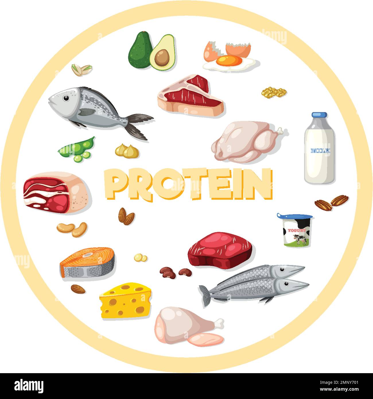
Humans can synthesize about half of these amino acids internally, but the remaining essential amino acids must come from dietary sources such as meat, eggs, dairy, or complementary plant combinations.
Formation of Peptide Bonds
Amino acids don't just float around in chains by accident; they're joined together through covalent peptide bonds. This connection forms via a dehydration synthesis reaction, also known as a condensation reaction.
During this process, the carboxyl group of one amino acid reacts with the amino group of an incoming amino acid. Water is released as a byproduct, and a strong peptide bond forms between the carbonyl carbon and the nitrogen atom.
When many amino acids link together sequentially, they form a polypeptide chain. The chain has a distinct directionality, starting at the N-terminus with a free amino group and ending at the C-terminus with a free carboxyl group. Ribosomes read messenger RNA instructions to stitch these monomers together in exact, predetermined sequences.
From Monomers to Three-Dimensional Structure
A linear chain of amino acids rarely functions on its own. To become an active protein, the polypeptide must fold into a precise three-dimensional shape. This folding occurs in four distinct hierarchical levels:
- Primary Structure: The exact linear sequence of amino acids linked by peptide bonds. Even a single substitution here can cause severe functional defects, as seen in sickle cell anemia where one valine replaces a glutamate in hemoglobin.
- Secondary Structure: Local folding patterns driven by hydrogen bonding along the backbone. This creates alpha helices (coil shapes) and beta pleated sheets (accordion folds).
- Tertiary Structure: The overall three-dimensional shape of a single polypeptide chain, driven by interactions between R-groups, including hydrogen bonds, ionic bonds, hydrophobic packing, and disulfide bridges.
- Quaternary Structure: The assembly of multiple polypeptide subunits into a functional protein complex, such as the four subunits that make up functional hemoglobin.
You can explore more about structural biology fundamentals through resources provided by the National Human Genome Research Institute.
Common Mistakes in Protein Synthesis and Folding
Cellular machinery operates with high fidelity, but errors do occur. Understanding these slip-ups highlights why precise amino acid sequences matter so much:
- Missense Mutations: A single DNA base change codes for a different amino acid, sometimes altering protein stability or active site geometry.
- Misfolding: Cellular stress or genetic flaws can cause proteins to fold incorrectly. Misfolded proteins often clump together into toxic aggregates, a hallmark of neurodegenerative conditions like Alzheimer's disease.
- Truncation: Premature stop codons cut polypeptide chains short, destroying function entirely.
Cells employ specialized proteins called chaperones to assist folding and tag irrecoverably damaged molecules for destruction via the proteasome.
Frequently Asked Questions
What is the difference between an amino acid and a peptide?
An amino acid is a single monomer unit containing an amino group, a carboxyl group, and an R-group. A peptide is a short chain of two or more amino acids linked by peptide bonds. Once a chain grows very long and folds into a functional shape, it is typically called a protein.
Why are there only 20 standard amino acids?
While prebiotic chemistry could produce many different organic molecules, evolutionary pressure selected a robust set of twenty amino acids that balance chemical diversity, stability, and metabolic cost. Organisms can occasionally incorporate non-standard amino acids like selenocysteine through specialized genetic recoding, but the core twenty handle the vast majority of biological tasks.
Can amino acids be used for energy instead of building proteins?
Yes. When dietary protein intake exceeds synthesis requirements, or during prolonged starvation, excess amino acids undergo deamination in the liver. Their nitrogen groups are converted to urea for excretion, while the remaining carbon skeletons enter cellular respiration pathways to generate ATP.
How do peptide bonds resist breakdown inside the cell?
Peptide bonds possess partial double-bond character due to resonance, making them rigid and chemically stable under normal physiological conditions. Cells rely on specific enzymes called proteases to break these bonds safely when proteins reach the end of their lifecycle.