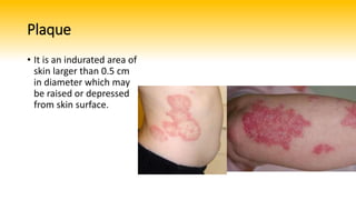
An induration wound isn't a distinct type of injury; it's a critical warning sign. Induration refers to the abnormal hardening or thickening of soft tissue surrounding a wound or injury site. When you notice firm, leathery, or rigid borders around a persistent sore, it signals deep-seated inflammation, unmanaged edema, or unresolved fibrosis. This tissue hardening chokes off capillary blood flow, starving the wound bed of oxygen and vital nutrients necessary for closure. Ignoring induration turns a slow-healing sore into a stalled, chronic wound vulnerable to deep tissue necrosis and systemic infection.
Understanding why tissue hardens requires looking beneath the skin's surface. Chronic wounds trap the body in a prolonged inflammatory loop. Instead of progressing smoothly from inflammation to proliferation and remodeling, the wound bed remains stuck. White blood cells flood the area relentlessly, releasing enzymes and cytokines that stimulate fibroblasts to deposit excessive collagen. This fibrous buildup creates the dense barrier known as induration. Without breaking this cycle, standard topical treatments fail because the local tissue architecture is too compromised to respond.
Core Causes of Tissue Hardening in Chronic Wounds
Induration develops when multiple pathological factors converge around a non-healing wound. Pinpointing the exact driver determines whether your treatment plan succeeds or fails.
- Persistent Biofilms and Infection: Bacteria don't just sit on the surface of a chronic sore; they organize into resilient biofilms. These microbial communities trigger a chronic immune onslaught, causing the surrounding skin to become rigid and inflamed.
- Venous Hypertension and Edema: In lower extremity ulcers, failing venous valves allow fluid to pool in the lower legs. Chronic fluid overload stretches the tissue, causing protein leakage into the interstitium and subsequent fibrosis or lipodermatosclerosis.
- Unrelieved Mechanical Pressure: Constant shear and pressure forces crush underlying microvessels. Ischemia sets in, and the body responds by laying down dense scar tissue to protect against further trauma.
- Uncontrolled Metabolic Disease: Conditions like diabetes impair angiogenesis and immune cell function. High blood glucose levels promote advanced glycation end-products that stiffen collagen matrixes across the body, making skin far more prone to induration.
Clinical Assessment: How to Evaluate Indurated Tissue
Accurate clinical evaluation goes beyond a simple visual check. You have to physically assess the tissue boundaries and density to gauge how deep the problem goes.
Palpation is your primary diagnostic tool. Gently press around the wound margin with clean gloved fingers. Healthy skin yields softly to pressure, whereas indurated tissue feels woody, firm, or board-like. Measure the exact radius of the hardened border outward from the wound edge, as this perimeter often reveals the true extent of subcutaneous inflammation or hidden undermining.
Differentiate clearly between standard perilesional erythema (redness) and true induration. Redness can stem from simple contact dermatitis or mild inflammation, but firmness indicates structural tissue remodeling and fibrosis. Clinicians should also assess local skin temperature, patient pain scores during palpation, and the presence of foul exudate or undermining. When induration extends rapidly or feels hot and boggy, suspect a deeper soft tissue infection or spreading cellulitis that requires immediate medical escalation.
Evidence-Based Treatment and Management Strategies
Treating a wound complicated by induration demands a multi-pronged approach. You cannot simply dress the top of the wound; you must address the physical barriers stiffening the surrounding tissue.
Debridement is non-negotiable. Surgical, sharp, or mechanical removal of necrotic tissue, slough, and fibrotic edges strips away the dead matrix fueling the inflammatory loop. Once the hostile tissue is cleared, fresh vascular beds can form. According to clinical guidance from the Wound Healing Society, aggressive management of bioburden and non-viable tissue is essential for jumpstarting stalled chronic wounds.
Compression therapy remains the gold standard for indurated venous leg ulcers. Applying graduated external pressure counteracts venous hypertension, drives fluid out of the interstitial spaces, and softens lipodermatosclerosis over weeks of consistent use. For pressure-induced induration, strict offloading using specialized boots, wheelchair cushions, or total contact casting is mandatory to relieve mechanical stress.
Moisture balance and advanced dressings support the newly liberated wound bed. Use antimicrobial or silver-impregnated dressings if bioburden is suspected, combined with non-adherent contact layers that protect fragile regenerating tissue. Furthermore, optimal nutrition—specifically adequate protein intake, vitamin C, and zinc—gives the body the raw materials needed to remodel fibrotic tissue into healthy extracellular matrix.
Frequently Asked Questions
What is the main difference between swelling and induration?
Swelling (edema) is soft, fluid-filled, and leaves an indentation when pressed. Induration is firm, dense, and rigid because it involves fibrous tissue buildup and chronic inflammation rather than just trapped water.
Can induration go away on its own?
Rarely. Because induration stems from chronic underlying pathology like poor circulation, persistent infection, or unmanaged pressure, it typically worsens unless the root cause is actively treated.
When does an indurated wound require emergency care?
Seek immediate medical attention if the induration spreads rapidly, the skin turns black or purple, you develop a high fever, or red streaks radiate away from the wound site. These are signs of severe spreading infection or tissue death.
How does compression therapy help soften hardened tissue?
Compression applies controlled external pressure that forces accumulated fluid out of the lower leg tissues, improves venous return, and reduces the chronic inflammation that drives fibrosis and skin thickening.