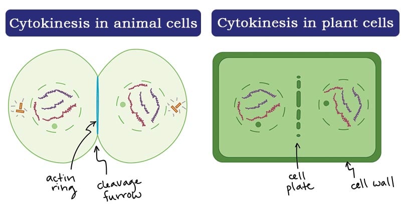
Cytokinesis is the final physical process of cell division that splits a single eukaryotic cell into two distinct daughter cells. It immediately follows mitosis, ensuring that each new cell receives a complete set of chromosomes and an appropriate share of cytoplasm and organelles. Without this critical concluding step, cells would become multinucleated anomalies unable to function normally.
The mechanics of cytokinesis differ significantly between animal and plant cells due to the presence of a rigid cell wall in plants. Animal cells rely on an actin-myosin contractile ring that pinches the plasma membrane inward, while plant cells construct a brand-new cell wall from the inside out using vesicles delivered by the phragmoplast. Understanding these pathways reveals how living organisms grow, heal, and maintain tissue integrity.
What is Cytokinesis and Why Does It Matter?
At its core, cytokinesis is the physical partitioning of the cellular contents following nuclear division. While mitosis ensures DNA is evenly separated, cytokinesis ensures the physical container splits correctly. If mitosis divides the library of genetic instructions, cytokinesis builds the walls for two separate libraries.
In multicellular organisms, precise control over this process prevents developmental disorders and uncontrolled tissue growth. Errors during this stage can lead to tetraploidy, where a cell retains double the normal amount of DNA. Such genetic instability is a hallmark of cancerous tumor formation. Researchers study these cellular checkpoints closely to understand how tissues maintain regular growth rates and normal chromosome counts.
For further reading on genetic stability and cell regulation, check resources from the National Institutes of Health.
The Animal Cell Pathway: The Contractile Ring
In animal cells, cytokinesis begins during anaphase or telophase of mitosis. The cell must determine precisely where to divide, a task managed by the mitotic spindle.
Spindle Midzone Signaling
Microtubules from the mitotic spindle signal to the cell cortex—the layer of protein filaments lying immediately beneath the plasma membrane—where the division plane should form. This positioning ensures that the genetic material is safely segregated on either side of the cleavage site before pinching begins.
Assembly of the Contractile Ring
Once the site is chosen, actin filaments and non-muscle myosin II molecules assemble into a ring directly beneath the plasma membrane. This contractile ring encircles the inner circumference of the cell perpendicular to the long axis of the mitotic spindle.
Cleavage Furrow Inward Pacing
As the myosin motors pull along the actin filaments, the ring tightens. This creates a visible indentation on the cell surface known as the cleavage furrow. The membrane pinches inward in a purse-string motion until the bridge narrows into a thin midbody structure. Tubulin and membrane-scission proteins then sever the connection, sealing the two new daughter cells completely.
The Plant Cell Pathway: The Phragmoplast and Cell Plate
Plant cells cannot pinch inward because a rigid cellulose cell wall surrounds them. Instead, they build a partition from the inside out.
Phragmoplast Formation
During late anaphase and telophase, microtubule structures called phragmoplasts assemble across the center of the division site. These structures act as railway tracks, guiding Golgi-derived vesicles carrying structural carbohydrates and enzymes toward the equatorial plane.
Cell Plate Assembly
The vesicles fuse together in the center of the cell, forming a flat, disk-like structure called the cell plate. As more vesicles arrive and fuse at the outer edges, the cell plate expands outward until it reaches the existing plasma membrane and sidewalls of the mother cell.
Maturation into a Cell Wall
Once the edges fuse with the parent cell wall, cellulose and pectin are deposited into the cell plate. This transforms the temporary partition into a permanent double cell wall separated by a middle lamella, fully dividing the two daughter cells.
Cytokinesis Versus Mitosis: Key Differences
People often confuse cytokinesis with mitosis because both occur during the broader cell cycle phase known as the M-phase. However, they are distinct processes with different responsibilities.
- Mitosis focuses exclusively on the nucleus and genetic material, dividing duplicated chromosomes into two identical sets across prophase, metaphase, anaphase, and telophase.
- Cytokinesis focuses on the cytoplasm, organelles, and plasma membrane, physically separating the cell body into two halves.
- Timing differs slightly as cytokinesis typically initiates late in anaphase or during telophase, overlapping with the final stages of nuclear division.
Common Mistakes and Misconceptions
Several common misunderstandings surround how cells divide. Clearing these up helps build an accurate picture of cellular mechanics.
- Assuming plant and animal cell division are identical: Many texts focus heavily on animal cleavage furrows, leading people to assume plant cells divide the same way. In reality, plant cells build an interior wall rather than pinching.
- Believing cytokinesis is part of mitosis: Mitosis strictly describes nuclear division. A cell can successfully complete mitosis without undergoing cytokinesis, which results in a single cell containing multiple nuclei.
- Thinking cytoplasm division is passive: The separation of the cytoplasm requires active molecular motors, specialized protein networks, and targeted vesicular transport, consuming chemical energy in the form of ATP.
For detailed educational breakdowns on molecular biology topics, visit the Nature Cell Biology Journal.
Frequently Asked Questions
What happens if cytokinesis fails?
If cytokinesis fails after mitosis, the cell ends up with twice the normal number of chromosomes, creating a binucleated or polyploid cell. While some specialized tissues like human liver cells tolerate polyploidy, uncontrolled failure in other tissues can lead to genetic abnormalities associated with cancer.
When does cytokinesis start in the cell cycle?
Cytokinesis typically begins during the anaphase or telophase stage of mitosis. The physical indentation or plate formation becomes visible shortly after sister chromatids have been pulled apart toward opposite poles of the cell.
Do all cells undergo cytokinesis immediately after mitosis?
No. Some cells undergo nuclear division repeatedly without splitting their cytoplasm, resulting in multinucleated cells called syncytia. Examples include skeletal muscle fibers in animals and certain fungi during rapid growth phases.
How do organelles like mitochondria get distributed during cytokinesis?
Organelles such as mitochondria and chloroplasts replicate independently throughout the cell cycle. When cytokinesis splits the cytoplasm, these organelles are distributed roughly between the two daughter cells, where they continue to multiply to meet metabolic demands.


