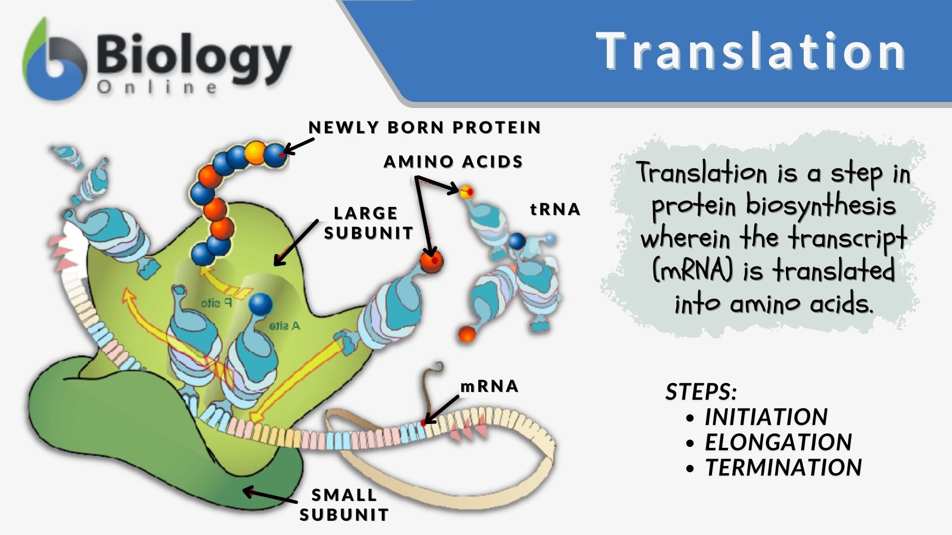
Gene expression isn't just about the protein-coding sequence of an mRNA molecule. Flanking this core region are the 5 prime and 3 prime untranslated regions, known as UTRs. While these segments don't code for amino acids, they control how much protein gets made, where the mRNA travels inside the cell, and how long the transcript survives before degradation.
When researchers or students look at messenger RNA, they often focus entirely on the open reading frame. Yet mutations or structural shifts in the non-coding flanks can completely alter cellular function. Understanding these regions clarifies post-transcriptional gene regulation in both healthy cells and disease states.
Anatomy and Mechanics of the 5 Prime UTR
The 5 prime UTR sits upstream of the protein-coding segment, spanning from the transcription start site to the initiation codon. Its primary job is governing translation initiation and ribosome recruitment.
In eukaryotes, the 5 prime UTR frequently contains complex secondary structures like hairpins and stem-loops. Ribosomes must scan through this region starting from the 7-methylguanosine cap to find the start codon. If the structure is too stable or tangled, scanning slows down, which typically reduces protein output. Conversely, streamlined sequences let ribosomes move quickly, boosting translation efficiency.
Key elements housed within the 5 prime UTR include:
- Upstream open reading frames: Short reading frames that initiate translation before the main protein start site, usually acting as roadblocks to dampen protein expression.
- Internal ribosome entry sites: Specialized structures allowing ribosomes to bind directly near or at the start codon, bypassing standard cap-dependent scanning, which is common during cellular stress or viral infection.
- Protein binding sites: Specific RNA motifs that bind translation repressor proteins to halt production when resources are scarce.
Function and Regulation within the 3 Prime UTR
Located downstream of the stop codon, the 3 prime UTR dictates mRNA stability, localization, and cellular half-life. Although it doesn't enter the ribosome's active decoding site, it serves as a bustling hub for microRNAs and RNA-binding proteins.
MicroRNAs bind to complementary sequences within the 3 prime UTR. When a microRNA locks onto its target, it recruits silencing complexes that either degrade the transcript or block ribosomes from translating it. This fine-tuning mechanism ensures that cells produce precise amounts of specific proteins rather than letting expression run unchecked.
Polyadenylation also plays a massive role here. Most eukaryotic mRNAs end with a poly-A tail. Shifts in the cleavage and polyadenylation sites can shorten or lengthen the 3 prime UTR, effectively stripping away or adding binding sites for regulatory factors. Longer regions tend to be more heavily regulated by microRNAs, while shortened versions often escape microRNA-mediated repression, leading to higher protein yields.
Real-World Impact on Disease and Cellular Stress
Dysregulation in untranslated regions triggers severe cellular consequences. Because these areas act as control switches, tiny genomic variations can drive pathology without changing a single amino acid in the final protein.
For instance, single nucleotide polymorphisms inside the 3 prime UTR can destroy a microRNA binding site. If a tumor suppressor gene loses its repressive microRNA site, it might express abnormally, or conversely, if a proto-oncogene gains stability through UTR mutations, cancer progression can accelerate.
Similarly, mutations in the 5 prime UTR can create aberrant upstream open reading frames. These rogue start sites trap scanning ribosomes, starving the main coding sequence of translation machinery and dropping essential protein levels drastically. Researchers studying genetic disorders now sequence these non-coding segments routinely, uncovering drivers of disease that coding-sequence analysis misses entirely.
Analyzing UTRs in Modern Research
Investigating these regions requires specialized techniques because standard whole-exome sequencing ignores non-coding flanks. Scientists rely on high-throughput approaches to map and quantify UTR activity across entire genomes.
- Polysome profiling: Separates mRNAs based on how many ribosomes are attached, revealing how efficiently specific 5 prime UTR variants translate in real time.
- Massively parallel reporter assays: Tests thousands of synthetic or mutated UTR sequences simultaneously in cell cultures to measure their direct impact on transcript stability and protein output.
- Cross-linking immunoprecipitation: Maps exact binding locations where regulatory proteins attach to untranslated RNA segments inside living cells.
Researchers utilizing these tools can engineer synthetic UTRs for biotechnology applications, such as optimizing mRNA vaccine stability and protein yield. Adjusting the length and GC content of the flanking regions allows scientists to fine-tune how long an injected transcript survives inside human tissue.
Frequently Asked Questions
Do prokaryotes have 5 and 3 UTRs?
Yes, prokaryotic mRNAs feature both 5 and 3 untranslated regions, though they are generally much shorter and structurally simpler than eukaryotic versions. The bacterial 5 prime UTR famously contains the Shine-Dalgarno sequence, which pairs with ribosomal RNA to position the start codon correctly.
Why are UTRs called untranslated if uORFs get translated?
While the overarching region is designated as untranslated because it sits outside the main protein-coding open reading frame, specific sub-elements like upstream open reading frames can direct the synthesis of small, often regulatory peptides. These short products rarely form functional structural proteins, keeping the primary label accurate for the bulk of the region.
How do UTRs affect mRNA localization?
Proteins and motor molecules recognize specific structural zipcodes embedded within the 3 prime UTR. These RNA-binding proteins escort the transcript to exact cellular destinations, such as synapses in neurons or the leading edge of a migrating cell, ensuring proteins are synthesized precisely where needed.
Can a mutation in a UTR cause cancer?
Yes, somatic mutations or structural rearrangements in UTRs can disrupt normal gene regulation. If a mutation eliminates a microRNA repression site on an oncogene, the cell may overproduce that protein, driving uncontrolled cell division.