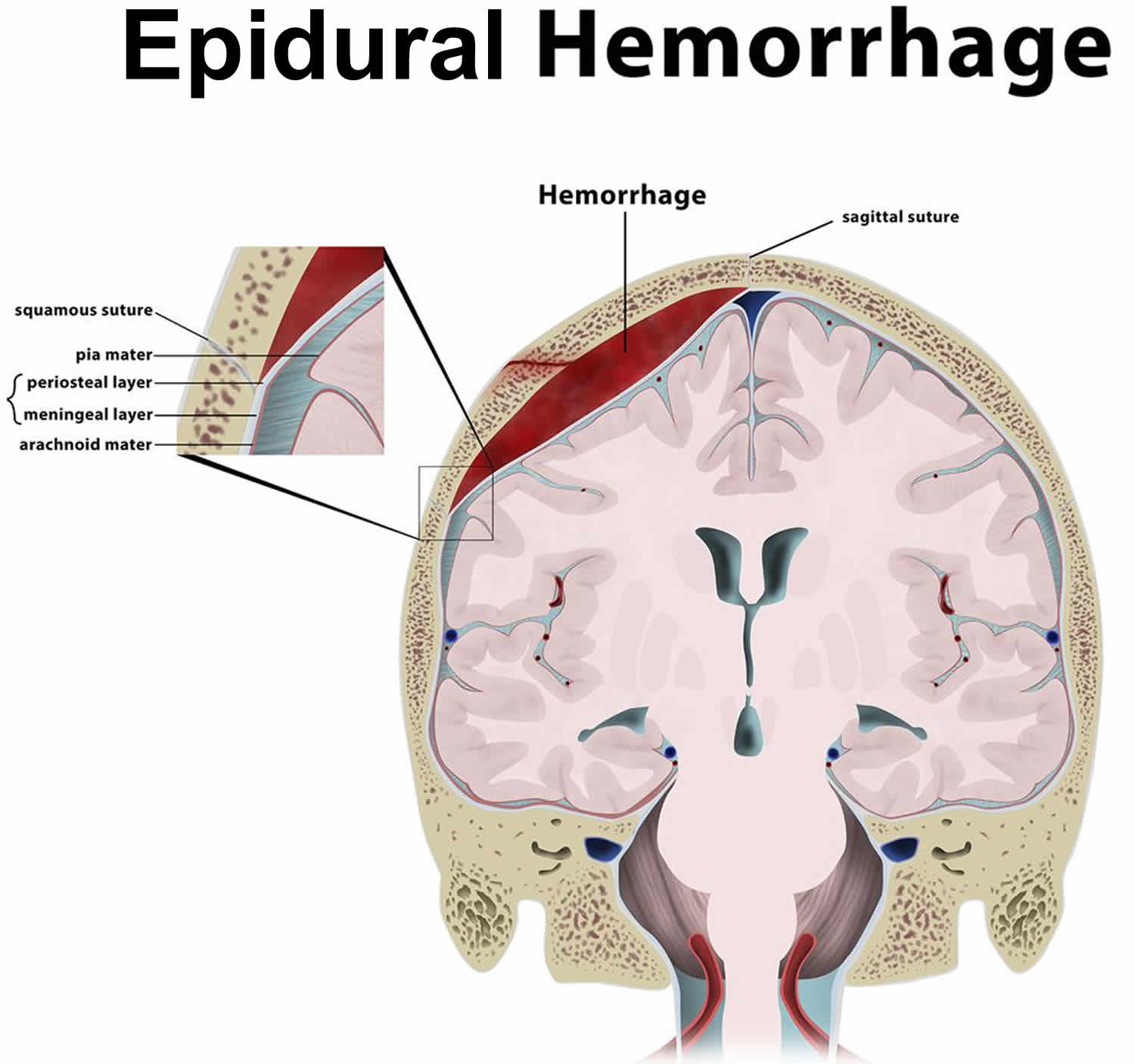
When a blood vessel tears, blood leaks into tissues where it doesn't belong. This pooling collection is called a hematoma. It's often confused with a basic bruise, but it's physically different because it involves a distinct pocket or mass of pooled blood rather than just surface discoloration.
You'll find these collections anywhere on the body, from superficial layers under the skin to deep intracranial spaces around the brain. Understanding how they form, when they're dangerous, and how doctors treat them helps you separate minor bumps from medical emergencies.
What Is a Hematoma and How Does It Form?
A hematoma occurs when trauma damages the walls of blood vessels—arteries, veins, or capillaries—causing blood to escape into surrounding tissue. Because blood pressure forces more fluid out of the damaged vessel, the blood accumulates and clots, forming a localized mass.
The body's natural response kicks in immediately to seal the vessel leak, but the trapped blood remains. Over time, surrounding tissue reabsorbs the pooled blood. This breakdown process changes the color of the affected area from dark red or purple to green, yellow, and finally brown before fading completely.
Unlike simple ecchymoses (common bruises), where blood leaks more diffusely into superficial tissues, hematomas create a palpable lump or swelling. They vary wildly in severity depending on their anatomical location and the type of blood vessel involved.
Primary Types of Hematomas
Doctors classify hematomas based on where they form in the body. The location dictates the urgency of treatment and the specific symptoms you'll experience.
Subcutaneous Hematomas
These develop directly underneath the skin. When you bump your shin hard against a coffee table, you get a subcutaneous hematoma. It's tender, swollen, and forms a visible lump. Most heal on their own as the body reabsorbs the blood.
Intramuscular Hematomas
When trauma damages muscle tissue deep beneath the skin, blood pools inside the muscle bundle. These are intensely painful because the rigid muscle fascia restricts swelling, increasing internal pressure. This pressure can compress nearby nerves and blood vessels, limiting mobility.
Intracranial Hematomas
These occur inside the skull and are medical emergencies. They include epidural, subdural, and intracerebral hematomas, which form between the skull and the brain or inside brain tissue itself. Causes usually involve severe head trauma, such as motor vehicle accidents or sports concussions. Symptoms may appear immediately or take days to manifest, making neurological monitoring critical after any major head injury.
Subnail Hematomas
A subnail hematoma forms directly underneath a fingernail or toenail, usually after dropping a heavy object on your hand or foot. The trapped blood creates intense, throbbing pressure beneath the rigid nail plate.
Common Causes and Risk Factors
Trauma is the leading trigger for these pooled blood collections, but it's not the only one.
- Blunt force trauma: Car crashes, falls, contact sports, and workplace accidents rupture blood vessels.
- Surgical complications: Procedures can inadvertently nick blood vessels, leading to postoperative collections.
- Anticoagulant medications: Blood thinners like warfarin, aspirin, or direct oral anticoagulants prevent normal clotting, making minor bumps turn into large hematomas easily.
- Bleeding disorders: Conditions like hemophilia impair the body's clotting cascade.
If you take blood thinners, even minor trauma requires close observation. For detailed guidance on medication management and bleeding risks, consult resources from the National Institutes of Health.
Recognizing Symptoms
Symptoms depend entirely on where the hematoma sits and how large it is. Superficial spots present clear physical clues, while deep tissue or internal collections hide their warning signs.
- Localized swelling: A firm, rubbery lump that feels tender or painful to the touch.
- Discoloration: Red, purple, blue, or black skin changes overlying the collection.
- Restricted movement: If a hematoma forms inside a quadricep or calf muscle, straightening or bending the limb hurts significantly.
- Numbness or tingling: Pressure on adjacent nerves creates a pins-and-needles sensation or localized weakness.
For intracranial hematomas, warning signs differ drastically. Watch for severe headaches, confusion, dizziness, vomiting, slurred speech, or unequal pupil size. These require emergency medical evaluation immediately. Learn more about emergency neurological signs through the Centers for Disease Control and Prevention.
Diagnostic and Treatment Approaches
Healthcare providers rely on physical evaluations and imaging to determine the exact size and depth of the blood collection.
Diagnosis
Doctors start with a physical exam to check the firmness, size, and tenderness of the lump. If they suspect internal bleeding or deep muscle involvement, they order imaging studies such as ultrasounds, CT scans, or MRIs. Blood tests help evaluate your overall clotting function, especially if you take medications that affect bleeding times.
Conservative Management
Small superficial or intramuscular hematomas often don't require surgery. Treatment follows standard injury protocols:
- Rest: Avoid using the injured muscle or limb to prevent further bleeding.
- Ice: Apply cold packs wrapped in a cloth for 15 to 20 minutes several times a day during the first 48 hours to constrict blood vessels.
- Compression: Use elastic bandages where appropriate to minimize spreading.
- Elevation: Raise the injured area above heart level to reduce fluid accumulation.
Surgical Intervention
Large hematomas, or those causing dangerous pressure on the brain, spinal cord, or major nerves, require medical drainage. A physician may use a needle to aspirate the fluid or perform a small surgical incision to evacuate the clot and stop active bleeding vessels. For painful subnail hematomas, doctors often perform "trephination"—melting a tiny hole in the nail plate with a heated device to release trapped pressure safely.
Frequently Asked Questions
Can I drain a hematoma at home?
No. Attempting to pierce, squeeze, or drain a hematoma at home introduces bacteria directly into the tissue, risking severe localized infections or sepsis. Always let a medical professional handle drainage procedures.
How long does it take for a hematoma to go away?
Small subcutaneous collections typically resolve within two to three weeks as the body reabsorbs the blood. Larger intramuscular hematomas can take several weeks or even months to disappear completely, depending on your age and overall circulation.
When should I see a doctor for a bruise or lump?
Seek medical evaluation if the lump continues growing, causes severe pain, creates numbness or muscle weakness, or appears on your head following an impact. You should also consult a physician if you take blood thinners and notice large, unexplained swelling.
What is the difference between a bruise and a hematoma?
A bruise is a flat discoloration caused by minor capillary leakage under the skin. A hematoma is a localized collection of blood that forms an actual palpable mass or pocket within tissues.



