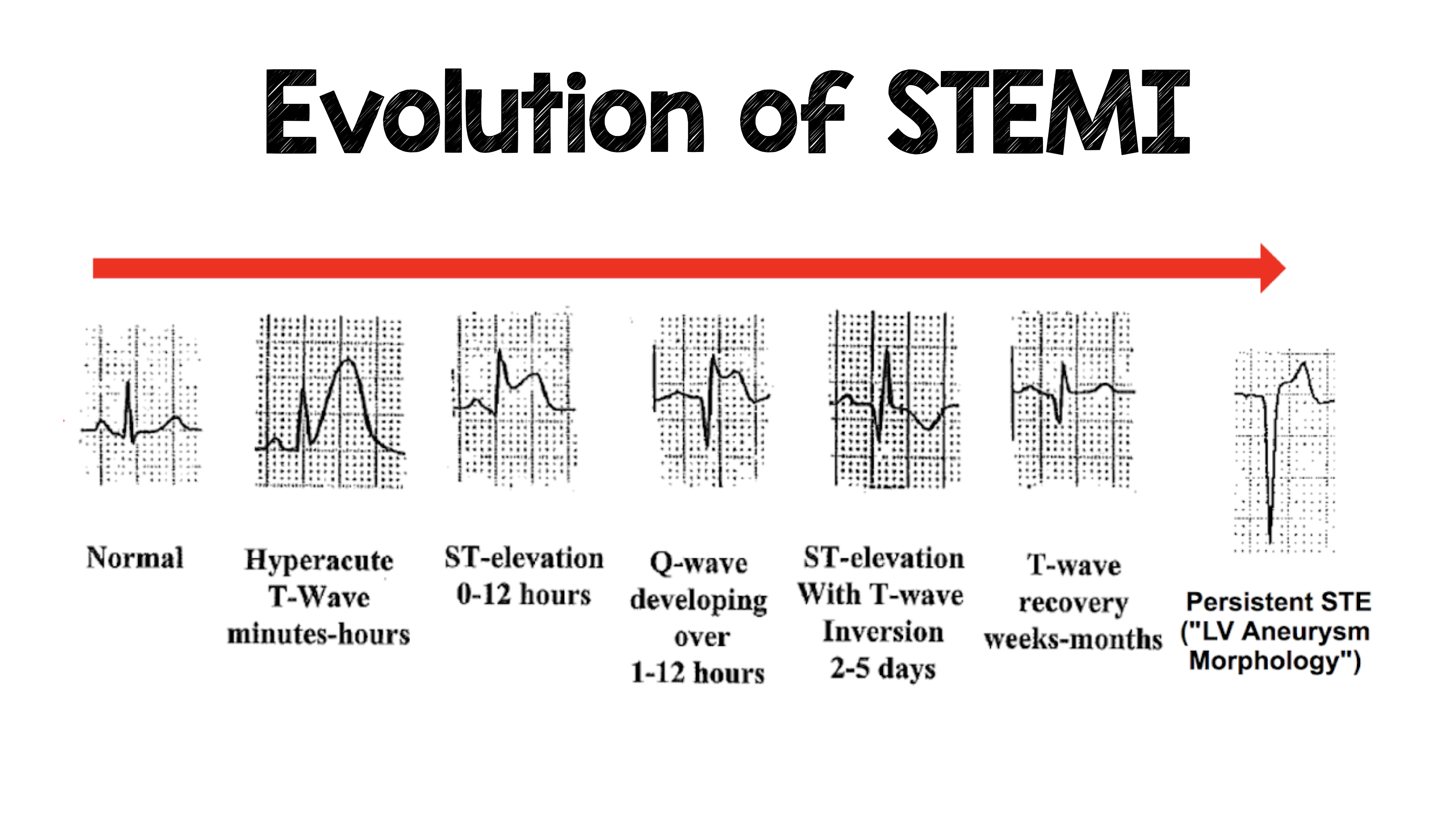
When a person experiences chest pain, emergency physicians and cardiologists immediately look for one critical finding on an electrocardiogram: ST-segment elevation. This single marker divides acute heart attacks into two distinct clinical categories: STEMI and NSTEMI. Knowing the difference between these two conditions isn't just an academic exercise; it dictates the speed, type, and urgency of lifesaving medical interventions.
Both STEMI and NSTEMI are forms of acute coronary syndrome, meaning they involve a sudden reduction or blockage of blood flow to the heart muscle. Yet their underlying pathology, ECG signatures, and immediate treatment pathways diverge significantly. Let's examine how these two myocardial infarctions differ in practice, diagnosis, and patient management.
Defining the Pathology: Occlusion vs Subtotal Blockage
To understand the core difference between the two events, you have to look at what's happening inside the coronary arteries.
A STEMI, or ST-elevation myocardial infarction, occurs when a coronary artery suffers a complete, 100% occlusion. Plaque ruptures, a blood clot forms rapidly over the rupture site, and blood flow halts entirely. Without immediate intervention, the heart muscle cells supplied by that vessel begin to die off rapidly within minutes.
An NSTEMI, or non-ST-elevation myocardial infarction, involves a severe but incomplete or intermittent blockage. The artery isn't entirely sealed off by the clot or plaque, so a trickle of blood still manages to pass through. While less abrupt in its total closure, tissue damage still occurs because the downstream heart muscle isn't receiving enough oxygen to meet its baseline metabolic demands.
According to guidelines from the American Heart Association, these distinct mechanical differences require entirely separate initial triage protocols in emergency departments.
Electrocardiogram and Biomarker Diagnostics
The primary tool used to separate these two events in the emergency room is the 12-lead electrocardiogram.
In a STEMI, the electrical current passing through the injured heart muscle creates a distinct visual signature on the ECG tracing: the ST segment lifts above the baseline. This classic " tombstone sign" tells the medical team that a major vessel is blocked right now and needs mechanical opening without delay.
In an NSTEMI, the ECG may show ST-segment depression, T-wave inversions, or sometimes no specific ischemic changes at all. Because the ECG doesn't show that classic elevation, doctors rely heavily on blood tests to confirm tissue damage. Specifically, they measure cardiac troponin levels, proteins that leak into the bloodstream when heart muscle cells suffer injury.
- STEMI Diagnosis: Confirmed primarily by characteristic ST-elevation on ECG, backed up by subsequent troponin elevation.
- NSTEMI Diagnosis: Confirmed by elevated troponin levels in the blood, combined with symptoms and non-elevated ECG patterns.
Treatment Pathways and Urgency
Because time equals muscle in cardiology, the speed of treatment varies between the two conditions.
For a STEMI, the goal is immediate mechanical reperfusion. Patients are rushed to the catheterization laboratory for a primary percutaneous coronary intervention, commonly called a PCI or angioplasty. A catheter is threaded through an artery in the wrist or groin to clear the clot and place a stent, opening the vessel back up. If a hospital with a catheterization lab isn't nearby, emergency teams administer clot-busting medications known as thrombolytics.
For an NSTEMI, the approach is urgent rather than emergent. Because the artery isn't 100% blocked, patients are typically stabilized first with medications like antiplatelets, anticoagulants, beta-blockers, and nitroglycerin. They still need a coronary angiogram and likely a stent, but the procedure is usually scheduled within 24 to 72 hours rather than immediately.
Common Mistakes in Patient Triage and Recovery
Misinterpreting or delaying treatment for acute coronary syndromes can lead to severe complications. Several common pitfalls often complicate care:
- Dismissing atypical symptoms: Women, older adults, and diabetic patients frequently present with subtle signs like nausea, shortness of breath, or fatigue rather than classic crushing chest pain.
- Waiting for lab results in a STEMI: Emergency teams don't wait for troponin blood test results to treat a suspected STEMI; the ECG alone is enough to trigger an emergency catheterization lab activation.
- Underestimating NSTEMI risk: Some patients assume an NSTEMI is "minor" because it isn't a STEMI. In reality, long-term mortality rates for NSTEMI catch up to STEMI over several years due to underlying multi-vessel coronary disease.
For more details on cardiovascular treatment standards, consult resources provided by the National Institutes of Health.
Frequently Asked Questions
Which heart attack type is more dangerous?
A STEMI is generally considered more immediately lethal because the complete blockage causes rapid, extensive damage to a large area of the heart muscle. However, NSTEMI patients often have more complex underlying coronary disease, leading to similar long-term risks.
Can an NSTEMI turn into a STEMI?
Yes. If a partial blockage in an NSTEMI progresses and a blood clot completely seals off the artery, the condition can evolve into a full STEMI. This is why close monitoring in a cardiac care unit is mandatory.
Do both conditions require stent placement?
Most patients with either condition will undergo a coronary angiogram. If significant blockages are found, cardiologists typically place stents during a PCI, or recommend bypass surgery for multi-vessel disease.
What are the shared risk factors for both?
Both conditions share identical root causes, including high blood pressure, high cholesterol, cigarette smoking, diabetes, physical inactivity, and a family history of premature heart disease.