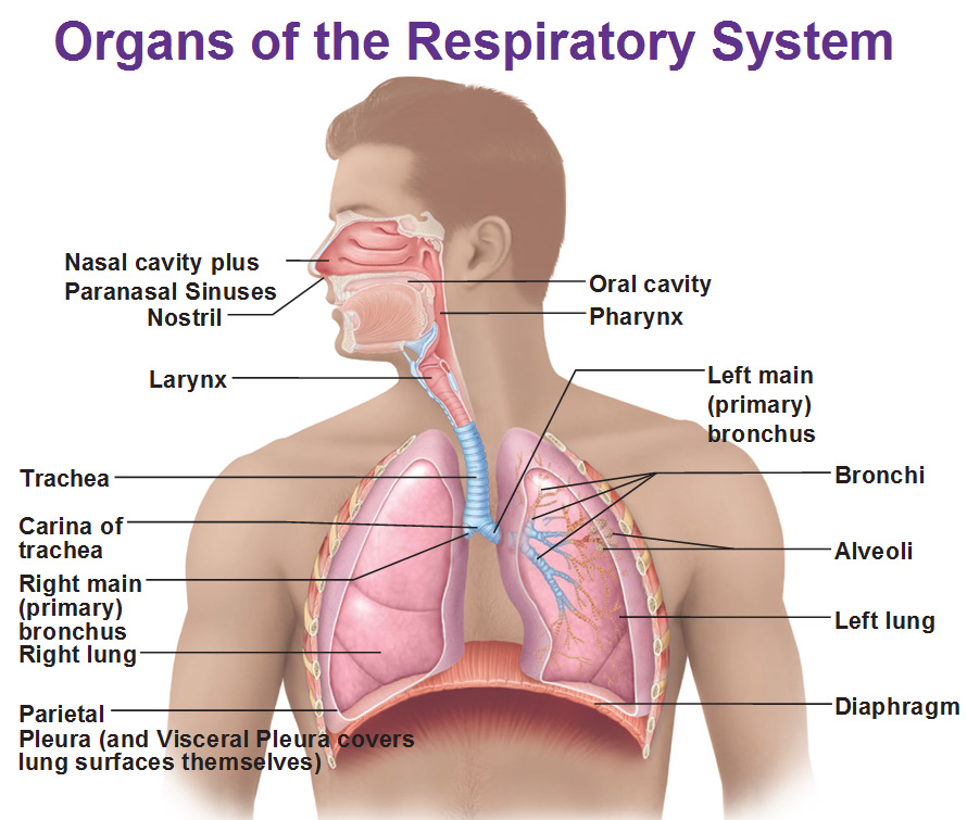
The human body requires a continuous supply of oxygen to generate cellular energy and eliminate metabolic waste products like carbon dioxide. The respiratory system manages this vital gas exchange through a coordinated sequence of physical and chemical processes that run automatically from the moment you are born. Understanding how air travels from the external environment into the deepest recesses of your lung tissue reveals an intricate network of specialized filters, muscular pumps, and microscopic membranes.
Upper Respiratory Tract Structure and Function
Air enters the body primarily through the nasal cavity, which acts as the initial processing plant for inhaled air. The nasal passages are lined with tiny, hair-like projections called cilia and specialized mucous membranes. As you inhale, these structures filter out particulate matter, dust, and airborne pathogens while warming and humidifying the incoming air to match core body temperature. This conditioning prevents delicate lower airway tissues from drying out and sustaining injury.
From the nasal cavity, air moves into the pharynx, a muscular conduit shared by both the digestive and respiratory tracts. A small, leaf-shaped flap of cartilage known as the epiglottis sits at the base of the throat. During swallowing, the epiglottis folds downward to seal off the larynx, directing food and liquids safely into the esophagus. When you breathe, the epiglottis stays upright, leaving the airway open so air can pass unimpeded into the trachea.
Lower Airway Mechanics and the Trachea
Once past the pharynx, air enters the trachea, commonly called the windpipe. The trachea is a rigid tube supported by a series of C-shaped rings of hyaline cartilage. These rings keep the airway open against changes in pressure during inhalation and exhalation, ensuring that the conduit doesn't collapse on itself.
The interior of the trachea features a lining of pseudostratified ciliated columnar epithelium. Glands embedded within this tissue secrete mucus that traps any remaining foreign particles. The coordinated, upward wave-like beating of the cilia—often referred to as the mucociliary escalator—sweeps trapped debris toward the pharynx, where it is swallowed or cleared by coughing.
At its lower end, the trachea branches into two primary bronchi:
- Right mainstem bronchus: Wider, shorter, and more vertical than the left, making it a common site for inhaled foreign objects to lodge.
- Left mainstem bronchus: Narrower and longer, angled slightly to accommodate the position of the heart in the thoracic cavity.
Bronchioles and Alveolar Architecture
Inside the lungs, the primary bronchi branch repeatedly into smaller secondary and tertiary bronchi, eventually narrowing into microscopic pathways called bronchioles. Unlike the trachea and large bronchi, bronchioles lack cartilage support. Instead, they feature bands of smooth muscle that contract or relax to regulate airflow resistance throughout the lungs.
Bronchioles terminate in clusters of tiny air sacs known as alveoli, which form the functional units of the respiratory system. An adult human lung contains roughly 300 to 500 million alveoli, creating an enormous surface area for gas exchange roughly equivalent to the size of a tennis court.
Each alveolus is wrapped in a dense network of pulmonary capillaries. The barrier separating the air inside an alveolus from the blood in the capillary is called the respiratory membrane. This membrane is exceptionally thin, consisting only of the alveolar epithelial cell layer, the capillary endothelial cell layer, and their fused basement membranes. This microscopic thickness allows respiratory gases to diffuse rapidly across the barrier without requiring active transport mechanisms.
Gas Exchange Physics and Partial Pressures
Gas exchange operates entirely through simple diffusion, driven by differences in partial pressures between the alveoli and the pulmonary capillary blood.
When you inhale, fresh air fills the alveoli, creating a high partial pressure of oxygen ($PO_2$) of roughly $104\text{ mmHg}$. Deoxygenated blood arriving from the right side of the heart via the pulmonary artery has a much lower oxygen partial pressure of about $40\text{ mmHg}$. Because gases move down their concentration gradients, oxygen molecules diffuse rapidly across the respiratory membrane from the alveolar gas phase into the deoxygenated blood plasma, binding efficiently to hemoglobin molecules inside red blood cells.
Simultaneously, the partial pressure of carbon dioxide ($PCO_2$) in the incoming deoxygenated blood is approximately $45\text{ mmHg}$, while the $PCO_2$ in the alveolar air is about $40\text{ mmHg}$. Carbon dioxide diffuses out of the blood plasma, across the respiratory membrane, and into the alveolar spaces to be expelled during exhalation.
For deeper context on how blood gas transport integrates with systemic circulation, refer to the overview provided by the National Institutes of Health.
Pulmonary Ventilation Muscles
Moving air in and out of the thoracic cavity requires active muscular work during inhalation and typically passive recoil during resting exhalation.
The primary muscle of respiration is the diaphragm, a dome-shaped sheet of skeletal muscle separating the thoracic and abdominal cavities. When you inhale, the diaphragm contracts and flattens downward, increasing the vertical dimension of the chest cavity. Simultaneously, the external intercostal muscles contract, pulling the rib cage upward and outward.
These coordinated movements increase thoracic volume and decrease intrapleural pressure below atmospheric pressure. Air rushes down this pressure gradient into the lungs. During quiet exhalation, the diaphragm and intercostal muscles relax, allowing the elastic recoil of the lung tissue and chest wall to decrease volume, raise internal pressure, and push air back out.
Neural Control and Regulation of Breathing
Breathing rate and depth are managed automatically by respiratory centers located in the brainstem, specifically the medulla oblongata and the pons. While you can hold your breath or alter your breathing pattern consciously for short periods, autonomic control overrides voluntary input to maintain blood gas homeostasis.
Central chemoreceptors located on the surface of the medulla oblongata monitor changes in the pH and carbon dioxide concentration of the cerebrospinal fluid. Because carbon dioxide readily crosses the blood-brain barrier, rising blood $CO_2$ levels lower cerebrospinal fluid pH, triggering the respiratory center to increase the rate and depth of ventilation. Peripheral chemoreceptors located in the carotid bodies and aortic bodies monitor arterial oxygen levels, carbon dioxide, and blood pH, providing rapid feedback during strenuous physical activity or hypoxic stress.
For clinical benchmarks on respiratory health diagnostics, you can review guidelines published by the American Lung Association.
Common Mistakes in Understanding Respiration
People often confuse cellular respiration with pulmonary ventilation. Pulmonary ventilation refers strictly to the mechanical movement of air into and out of the lungs. External respiration involves the exchange of gases between the alveoli and the pulmonary capillaries. Internal respiration describes gas exchange between systemic capillaries and active tissue cells throughout the body. Cellular respiration is an entirely different metabolic process occurring within cell mitochondria to produce adenosine triphosphate using oxygen.
Another frequent misunderstanding involves the mechanism of oxygen transport. While a small amount of oxygen dissolves directly into blood plasma, roughly ninety-eight percent of oxygen is carried through the circulatory system bound to hemoglobin proteins inside erythrocytes. Carbon dioxide is transported simultaneously via three distinct methods: dissolved in plasma, bound to hemoglobin carbamino complexes, and converted into bicarbonate ions for safe buffering in the bloodstream.
Frequently Asked Questions
What stops food from entering the trachea during swallowing?
The epiglottis functions as a flexible flap of cartilage at the base of the tongue. When you swallow, laryngeal muscles pull the larynx upward while the epiglottis folds backward over the opening of the airway, sealing off the trachea and diverting food into the esophagus.
Why is the right lung larger than the left lung?
The human heart sits slightly to the left of the body's midline in the thoracic cavity. To accommodate the cardiac notch and the apex of the heart, the left lung features only two lobes and a smaller overall volume, whereas the right lung contains three distinct lobes.
How does high altitude affect gas exchange?
At higher altitudes, atmospheric pressure drops, lowering the partial pressure of oxygen in the ambient air. This creates a smaller pressure gradient between the alveoli and the blood, reducing the efficiency of oxygen diffusion and causing symptoms of hypoxia until physiological adaptations occur.
What causes a stitch in the side during exercise?
A side stitch, or exercise-related transient abdominal pain, typically occurs when repetitive jarring movements during running pull on the ligaments connecting the diaphragm to internal organs, transiently reducing local blood supply to the respiratory muscles.