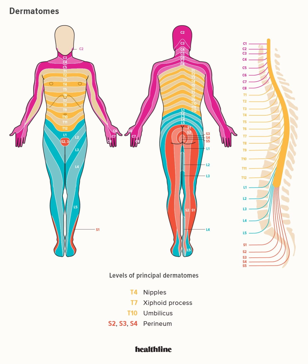
When you feel a patch of numbness on your shin or a sharp, shooting pain down the outside of your calf, your nervous system is sending a precise anatomical signal. Dermatomes of the lower leg are specific strips of skin supplied by sensory nerve fibers from a single spinal nerve root. Mapping these regions allows clinicians to trace sensory changes directly back to the spine. If you've ever wondered why a slipped disc in your lower back makes your big toe numb, the answer lies in the overlapping layout of these nerve pathways.
Understanding this map isn't just for neurosurgeons or physical therapists. Whether you're tracking an injury or studying human physiology, knowing how your skin senses touch and temperature relative to your spinal segments clarifies how pain travels. Let's break down the exact pathways, landmark regions, and clinical testing methods that define lower extremity sensation.
What is a Dermatome?
A dermatome is an area of skin where all the sensory input feeds into a single dorsal root of a spinal nerve. Along the human body, these segments stack like stacked rings or vertical stripes. While the trunk forms neat horizontal bands, the limbs twist and rotate during embryonic development. This developmental shift stretches the dermatomes into longitudinal stripes down the legs.
The lower limb relies primarily on spinal levels L1 through S2, with sacral levels S3 through S5 wrapping up the perineal region. Because adjacent nerve roots overlap significantly, losing the function of a single root rarely causes total numbness in a clean geometric patch. Instead, it creates a zone of diminished sensation—known as hypoesthesia—centered on that nerve's primary territory.
Mapping the Lower Leg Dermatomes
The lumbar and sacral spinal nerves divide the lower extremities into distinct topographical zones. Moving from the hip down to the toes, each segment claims a specific real estate of skin.
Lumbar Innervation (L1 to L5)
The lumbar spine governs the front, inside, and outer corridors of the lower limbs:
- L1: Covers the inguinal crease and the upper anterior region of the thigh.
- L2: Wraps across the middle and anterior-medial thigh.
- L3: Descends to cover the medial femoral condyle and the region right around the medial knee.
- L4: Travels down the medial leg, crosses the medial malleolus (the bony bump on the inside of your ankle), and extends toward the medial edge of the foot.
- L5: Covers the lateral aspect of the lower leg, sweeps across the dorsum (top) of the foot, and targets the first web space between the big toe and the second toe.
Sacral Innervation (S1 to S5)
The sacral nerves generally take charge of the back of the leg, the heel, and the sole:
- S1: Supplies the lateral foot, the small toe, the heel, and the posterior calf.
- S2: Covers the middle of the popliteal fossa (the back of the knee) and parts of the posterior thigh.
- S3, S4, and S5: Supply the perineal region, gluteal crease, and perianal skin, forming the classic "saddle anesthesia" distribution if injured.
For more detailed anatomical charts and clinical reference guides, you can explore the National Institutes of Health StatPearls library.
Clinical Significance: Why Dermatomes Matter
When a disc herniates in the lumbar spine, it typically bulges backward and pinches an exiting nerve root. This condition, called radiculopathy or colloquially known as sciatica, doesn't just hurt everywhere—it creates a targeted pattern of symptoms.
A clinician uses dermatomal maps to reverse-engineer back pain. If a patient reports tingling exclusively along the top of the foot and the big toe, the target suspicion falls squarely on the L5 nerve root. If the outer edge of the foot and the heel feel numb, the S1 root is usually the culprit.
Common Mistakes in Sensory Testing
- Ignoring overlapping borders: Expecting textbook, hard-line boundaries between nerve territories leads to diagnostic errors because adjacent nerves share innervation zones.
- Testing with heavy pressure: Heavy pressure recruits deep touch receptors rather than superficial cutaneous nerves. Light touch requires minimal skin deflection.
- Failing to compare sides: Always test matching areas on both legs to establish the patient's individual baseline.
How Clinicians Test Lower Leg Sensation
Neurological exams rely on two primary sensory modalities to evaluate lower leg dermatomes:
- Light Touch: The examiner uses a wisp of cotton or a soft monofilament, gently brushing it across specific skin landmarks while the patient's eyes are closed.
- Pinprick Sensation: A sterile safety pin or specialized neurological wheel tests sharp pain perception, helping differentiate pathway blocks.
You can review standard examination protocols through resources provided by OrthoFixar. Paired with deep tendon reflex checks—like tapping the patellar tendon for L4 or the Achilles tendon for S1—these tests offer a window into spinal cord health.
Frequently Asked Questions
Why do dermatome maps vary slightly between medical textbooks?
Textbook maps show idealized, simplified boundaries. In human anatomy, extensive overlap between adjacent spinal nerves means sensory zones blend into one another rather than meeting at sharp borders.
What is the difference between a dermatome and a peripheral nerve distribution?
A dermatome represents the area supplied by a single spinal nerve root before it bundles into peripheral nerves. A peripheral nerve (like the sciatic or peroneal nerve) contains fibers from multiple spinal roots bundled together, resulting in a different pattern of numbness if injured.
Can a single nerve root injury cause muscle weakness as well as numbness?
Yes. Spinal nerve roots carry both sensory and motor fibers. An L5 compression can cause numbness on the top of the foot alongside weakness when lifting the front of the foot upward.
How do doctors test the S1 dermatome specifically?
Clinicians test S1 sensation by checking the skin along the lateral and plantar aspects of the foot and the little toe, often paired with checking the ankle reflex.



