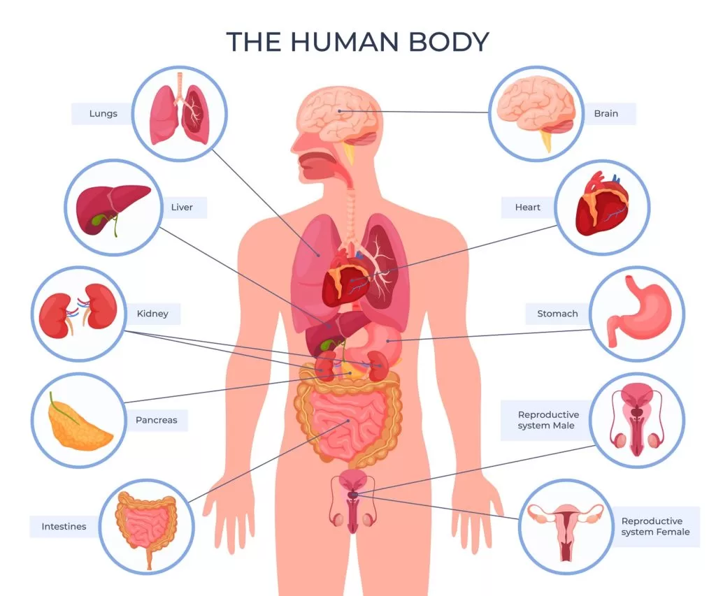
Understanding how the human body works starts with mapping out its core components. A human body parts diagram serves as a visual index for the skeletal, muscular, nervous, and circulatory frameworks, translating flat textbook pages into a functional blueprint of your physical self. Whether you're studying for an introductory biology class or simply trying to figure out where your pancreas actually sits, breaking the body down by its regional regions and physiological systems makes complex medical concepts click.
Anatomy isn't just a collection of isolated organs; it's an interdependent network where structure dictates function. When you look at an anatomical chart, you're observing millions of years of evolutionary engineering designed to keep your cells oxygenated, your posture upright, and your thoughts processing at lightning speed. Let's map out how these layers fit together from head to toe.
Major Anatomical Regions of the Body
Before diving into individual organs, clinical references divide the physical form into distinct geographical zones. This regional approach helps medical professionals and students pinpoint symptoms and locate structures quickly.
- The Head and Neck Region: Houses the brain, sensory organs (eyes, ears, nose, tongue), and the upper sections of the respiratory and digestive tracts. The skull protects the central processing unit, while the cervical vertebrae allow for a wide range of motion.
- The Thoracic (Chest) Region: Enclosed by the rib cage, sternum, and thoracic spine. This cavity shields vital life-support equipment, specifically the heart and lungs, keeping them safe from external impacts.
- The Abdominopelvic Region: A large cavity stretching from the diaphragm down to the floor of the pelvis. It contains the stomach, liver, intestines, kidneys, and reproductive organs without the rigid bone protection found in the chest, relying instead on muscular abdominal walls.
- The Upper and Lower Limbs: The arms and legs provide mobility and dexterity. Each limb is structured around a strong proximal bone (like the humerus or femur) that tapers down into intricate networks of smaller bones in the hands and feet.
The Core Physiological Systems
When examining a detailed body diagram, you'll notice that organs are grouped into systems based on shared objectives. There are twelve primary systems that keep a human alive and functioning.
Skeletal and Muscular Frameworks
The skeletal system consists of 206 bones in an adult human. It provides a rigid structural framework, anchors your muscles, and protects soft internal organs. For example, your cranium shields your brain, while your rib cage protects your heart and lungs.
Working hand-in-hand with the bones is the muscular system. Muscles contract and relax to pull on skeletal levers, creating movement. Skeletal muscles are voluntary, meaning you control them, while smooth and cardiac muscles operate automatically to manage digestion and blood circulation.
Circulatory and Respiratory Networks
Your cells need a constant supply of oxygen and nutrients to survive. The cardiovascular system handles this delivery using the heart as a central pump, driving blood through a vast network of arteries, veins, and capillaries.
The respiratory system feeds fresh oxygen into that blood. When you inhale, air travels down the trachea into the lungs, where oxygen crosses microscopic membranes into the bloodstream while carbon dioxide is expelled. You can explore interactive models of these networks via InnerBody Research to see how blood vessels weave through respiratory tissues.
Control and Communication Centers
The nervous system acts as the body's electrical grid. It includes the brain, spinal cord, and peripheral nerves. It processes sensory input from your environment, triggers muscle reactions, and regulates unconscious processes like blinking or swallowing.
Working alongside the nervous system is the endocrine system. Instead of electrical impulses, it uses chemical messengers called hormones. Glands such as the thyroid, adrenal glands, and pancreas secrete hormones directly into the bloodstream to regulate metabolism, growth, and stress responses.
Digestion, Filtration, and Defense
Processing fuel and clearing out waste involves several specialized systems working in sequence:
- Digestive System: Breaks down food mechanically and chemically from the mouth down through the esophagus, stomach, small intestine, and large intestine, extracting nutrients and expelling solid waste.
- Urinary System: Kidneys filter waste products and excess water from the blood, producing urine which is stored in the bladder before elimination.
- Lymphatic and Immune System: A drainage and defense network of lymph nodes and vessels that clears tissue fluids and hunts down pathogens.
How to Read an Anatomical Diagram Effectively
Looking at a complex medical chart can feel overwhelming if you don't know what you're looking at. Diagrams use specific planes and spatial orientations to make three-dimensional biology readable on a flat surface.
Planes of section divide the body into standard views. A coronal (frontal) slice splits the body into front and back halves. A sagittal slice divides it into left and right sides. A transverse slice cuts horizontally, separating the upper body from the lower body.
When studying a diagram, always locate the anterior (front) and posterior (back) markers first. This orientation prevents you from confusing internal organs that might look similar from different angles. For official anatomical terminology standards, you can review references provided by the National Institutes of Health.
Common Misconceptions About Internal Anatomy
Pop culture and casual conversation often misplace key organs. Clearing up these errors helps you read a body diagram with greater accuracy.
Many people point to their chest when talking about the stomach, but the stomach actually sits much lower, tucked just below the rib cage in the upper left section of the abdomen. The heart isn't sitting squarely on the left side of your chest either; it sits nearly dead center behind the sternum, with its bottom tip tilted slightly to the left.
Another frequent mix-up involves the kidneys. People often think they sit right at hip level in the lower back. In reality, the kidneys sit higher up under the lower ribs, protected by the lower part of the rib cage, with the right kidney sitting slightly lower than the left to accommodate the liver.
Frequently Asked Questions
What is the difference between anatomy and physiology?
Anatomy focuses on the physical structure and location of body parts, while physiology examines how those parts function and work together to sustain life.
Why do medical diagrams use Latin terms?
Anatomical naming conventions rely heavily on Latin and Greek roots to maintain a universal language that medical professionals worldwide can understand, regardless of their native tongue.
How many organ systems are in the human body?
Standard medical education generally categorizes the human body into twelve major systems, ranging from the skeletal and muscular systems to the integumentary (skin) and reproductive systems.
Can I view interactive human anatomy online for free?
Yes, digital platforms like the MedlinePlus Anatomy Resources offer verified, interactive diagrams and educational tools for students and patients.



