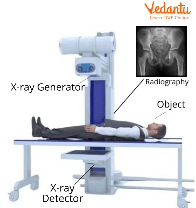
A hand X-ray is a quick imaging test that lets medical professionals inspect the 27 bones, numerous joints, and surrounding soft tissues of your hand and wrist. If you've ever fallen on an outstretched hand, felt persistent joint stiffness, or noticed swelling after a minor sports mishap, a clinician will likely order this exact diagnostic scan. It captures detailed radiographs in minutes, helping teams pinpoint fractures, track bone aging, and identify chronic joint deterioration without invasive procedures.
The procedure itself takes very little time and requires minimal preparation. You'll sit next to an X-ray machine while a technician positions your hand flat on a specialized plate in a few different angles—usually a standard dorsopalmar (back-to-front) view and an oblique angle. These varied projections ensure no small hairline fracture or overlapping bone gets hidden from view. Radiation exposure is extremely low, making it a safe frontline tool for patients of all ages, from children with growthplate concerns to older adults experiencing joint pain.
What a Hand X-Ray Shows
When a radiologist examines your hand radiographs, they look at several anatomical components simultaneously. Bone density and cortical thickness reveal how robust your skeletal framework is, which helps screen for conditions like osteoporosis. Joint spaces appear as dark gaps between bright white bones; a narrowing of these gaps often signals cartilage loss from wear and tear.
Beyond bones, the images capture alignment anomalies and foreign objects lodged under the skin, such as glass splinters or metal fragments. While X-rays primarily highlight hard structures, soft tissue swelling shows up as a hazy gray shadow around the joints, alerting your care team to localized inflammation or acute trauma.
Key Anatomical Structures Visible
- Phalanges: The fourteen finger bones spanning your proximal, middle, and distal segments.
- Metacarpals: The five long bones forming the palm of your hand.
- Carpals: The eight compact wrist bones arranged in two neat rows.
- Joint spaces: The protective cartilage gaps cushioning bone ends.
Common Medical Indications
Doctors order hand X-rays to investigate a wide array of symptoms and injuries. Acute trauma is the most frequent reason; sports injuries, workplace accidents, and clumsy falls frequently cause fractures in the distal phalanges or metacarpals. Radiographs confirm whether a broken bone is displaced, which dictates whether you need a simple splint or surgical fixation.
Chronic inflammatory conditions also prompt regular imaging. For example, rheumatoid arthritis creates symmetric joint damage, bone erosions, and soft tissue swelling that show up clearly over time. Osteoarthritis, by contrast, typically presents as asymmetrical joint space narrowing, bone spurs known as osteophytes, and subchondral sclerosis at the base of the thumb or finger joints.
Signs That Warrant an X-Ray
- Persistent swelling or bruising that doesn't improve after three days of rest.
- Inability to straighten or bend a finger fully following an impact.
- Chronic morning stiffness lasting longer than thirty minutes in multiple hand joints.
- Visible deformity, numbness, or a suspected foreign body embedded under the skin.
Limitations of Standard Hand Radiographs
While hand X-rays are invaluable, they don't capture everything. Because X-ray beams pass easily through soft tissues, standard films cannot visualize ligaments, tendons, or cartilage directly. If you suffer from a torn tendon, a strained collateral ligament, or early-stage carpal tunnel syndrome, your hand X-ray will typically look completely normal.
To investigate soft tissue damage, doctors rely on advanced imaging techniques. An MRI scan provides detailed views of muscles, tendons, and nerve pathways, while musculoskeletal ultrasound helps evaluate dynamic joint movement and superficial tendon tears in real time. Knowing these limitations prevents unnecessary delays when dealing with soft tissue injuries.
Preparing for Your Appointment
Preparation for a routine hand X-ray is straightforward. You don't need to fast or follow special dietary restrictions. Before the technician takes the images, you'll need to remove all rings, bracelets, and watches from the hand and wrist being scanned, as metal objects block X-ray beams and obscure underlying bone structures.
Let the radiology technician know if there's any chance you might be pregnant. While the radiation dose directed at your hand is extremely low and scattered far from your abdomen, clinics take standard precautions to minimize exposure. Patients with previous surgical hardware, such as pins or plates from an old fracture repair, should mention this so the radiologist can interpret the healing bone architecture accurately. You can read more about radiology safety standards at the RadiologyInfo portal.
Frequently Asked Questions
Does a hand X-ray hurt?
No, the test is entirely painless. The technician will position your hand on the digital plate, but you won't feel any physical discomfort during the exposure.
How long does the procedure take?
The actual imaging process takes less than five minutes. From check-in to walking out the door, plan for about fifteen to twenty minutes total.
Can a hand X-ray detect arthritis?
Yes, standard radiographs show classic signs of arthritis, including joint space narrowing, bone spurs, and bone-on-bone rubbing. However, very early cartilage inflammation might require an MRI for definitive detection.
Are children allowed to have hand X-rays?
Yes, pediatric hand X-rays are common for evaluating sports injuries or assessing bone age during growth and development. Technicians use specialized shielding to keep radiation exposure as low as reasonably achievable. Learn more about pediatric care guidelines through the American Academy of Pediatrics.



