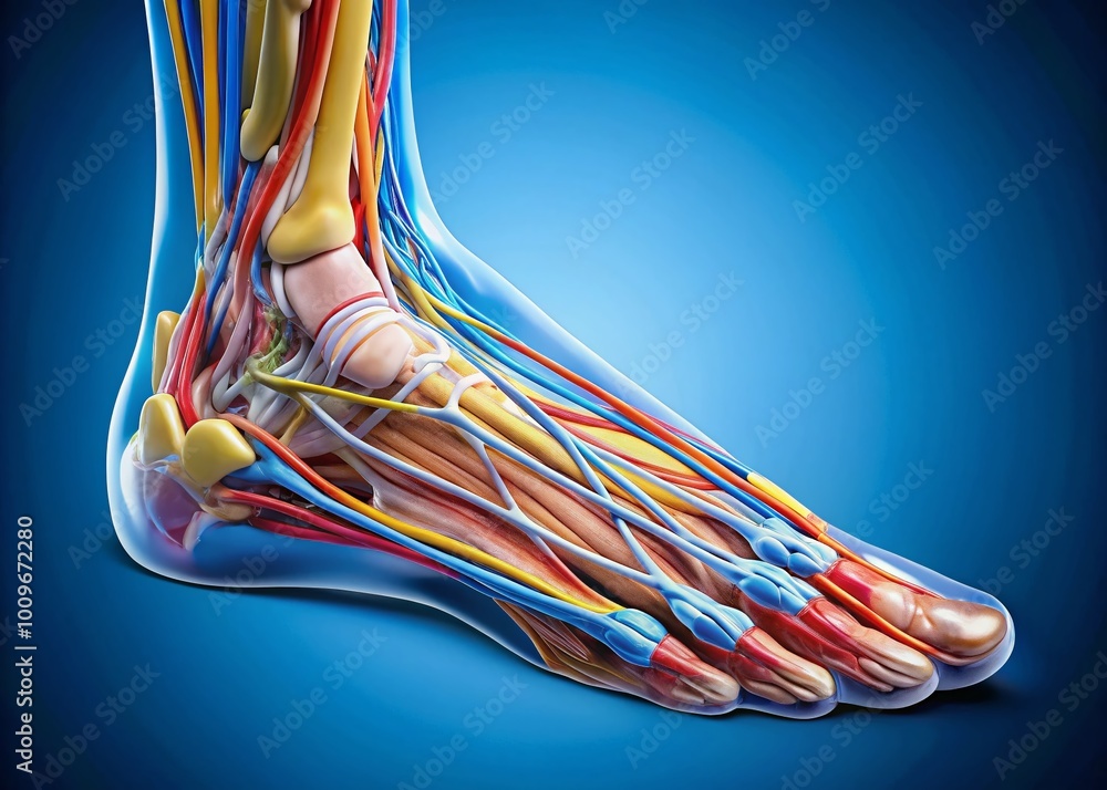
Your foot absorbs roughly 300 tons of cumulative force during a standard one-hour walk. Comprising 26 bones, 33 joints, and over a hundred muscles, tendons, and ligaments, this complex structure converts dynamic impact into propulsion while maintaining balance across unpredictable terrain.
Understanding foot anatomy requires looking past a surface-level visual diagram to examine how structural regions, intrinsic mechanics, and force distribution interact during movement. This guide breaks down the structural architecture of the human foot, joint mechanics during the gait cycle, common clinical pathologies, and practical assessment protocols for clinical and athletic performance.
Regional Anatomy and Bony Architecture
The human foot splits structurally into three distinct regions: the hindfoot, midfoot, and forefoot. Each division plays a specialized role in absorbing ground reaction forces and driving the body forward.
Hindfoot: The Foundation and Shock Absorber
Formed by the talus and calcaneus, the hindfoot forms the primary landing zone during heel strike.
- The Calcaneus: As the largest bone in the foot, the calcaneus features a prominent tuberosity that receives initial ground contact and anchors the Achilles tendon. Its dense trabecular network handles high compressive loads.
- The Talus: Sitting directly above the calcaneus, the talus forms the inferior component of the ankle joint (talocrural articulation). It lacks direct muscular attachments, relying instead on capsule and ligament stability.
- The Subtalar Joint: Positioned between the talus and calcaneus, this articulation allows triplanar motion consisting of inversion, eversion, abduction, and adduction. It acts as a torque converter, transforming tibial rotation into foot pronation and supination.
Midfoot: The Flexible Bridge
The midfoot consists of five tarsal bones: the navicular, cuboid, and medial, intermediate, and lateral cuneiforms. These bones form a rigid yet adaptable vault known as the transverse and medial longitudinal arches.
- The Navicular: Positioned on the medial side, the navicular articulates with the talus head. Its tuberosity serves as the primary insertion point for the tibialis posterior tendon, which maintains arch height.
- The Cuboid: Located laterally, the cuboid articulates with the calcaneus and fourth and fifth metatarsals, guiding the lateral column of the foot.
- Arch Mechanics: The midfoot functions as a shock absorber. During weight-bearing, these bones articulate to flatten slightly, dispersing energy before locking into a rigid lever for push-off.
Forefoot: The Propulsive Lever
Consisting of five metatarsals and 14 phalanges, the forefoot handles final load transfer and terminal propulsion.
- Metatarsal Heads: The first and fifth metatarsal heads bear the highest plantar pressures during the terminal stance phase.
- Sesamoid Bones: Two small sesamoids sit beneath the first metatarsophalangeal joint within the tendon of the flexor hallucis brevis. They protect the tendon, increase mechanical leverage for the great toe, and distribute weight during toe-off.
- Phalanges: The big toe (hallux) contains two phalanges, while the lateral four toes contain three each, providing balance adjustments during the final phase of the stride.
Articulations and Joint Mechanics
The joints of the foot operate through coordinated sequences. According to the American Orthopaedic Foot & Ankle Society, normal foot function depends on the precise timing of joint unlocking and locking throughout movement.
The Talocrural Joint
This uniaxial modified hinge joint permits dorsiflexion and plantarflexion. The stability of the mortise—formed by the distal tibia, medial malleolus, and lateral malleolus of the fibula—restricts side-to-side motion when the ankle is in dorsiflexion.
The Midtarsal Joint (Chopart Joint)
Comprising the talonavicular and calcaneocuboid joints, the midtarsal complex connects the hindfoot to the midfoot. When the subtalar joint pronates, the axes of the midtarsal joint become parallel, making the foot flexible to absorb uneven surfaces. When the subtalar joint supinates, these axes diverge, locking the midfoot into a rigid lever.
Metatarsophalangeal (MTP) Joints
The first MTP joint is critical for normal walking. It requires at least 65 degrees of dorsiflexion during terminal stance to allow the body to roll forward over the toes—a mechanism known as the windlass mechanism, where tension in the plantar fascia elevates the medial longitudinal arch as the toes extend.
Biomechanics of the Gait Cycle
During walking and running, the foot experiences distinct phases of ground contact and force transmission.
- Initial Contact: The heel strikes the ground slightly inverted. The subtalar joint begins to pronate, unlocking the midfoot to absorb impact and adapt to the ground surface.
- Midstance: Body weight passes directly over the supporting limb. The tibia rotates externally over the fixed foot, driving subtalar supination and locking the midfoot bones together.
- Terminal Stance: The heel rises off the ground. The calf muscles contract concentrically, while the plantar fascia tightens via the windlass mechanism, transforming the foot into a rigid lever for propulsion.
- Swing Phase: The ankle dorsiflexes to clear the toes as the limb swings forward for the next step.
Common Clinical Pathologies and Localized Pain
Structural abnormalities or excessive biomechanical stress frequently manifest in specific regions of the foot.
Plantar Fasciitis
Inflammation or degenerative tearing of the plantar fascia typically originates at its medial insertion on the calcaneal tuberosity. Patients experience sharp pain during the first steps in the morning or after prolonged rest, as the fascia shortens and tightens while unloaded.
Hallux Valgus (Bunions)
This progressive deformity involves lateral deviation of the proximal phalanx of the great toe and medial deviation of the first metatarsal bone. This malalignment alters weight distribution across the forefoot, often leading to bursitis over the medial eminence and compensatory overload of the lesser metatarsals.
Lateral Ankle Sprains
Inversion injuries during plantarflexion damage the lateral ligament complex. The anterior talofibular ligament tears first, followed frequently by the calcaneofibular ligament. Chronic instability can alter subtalar joint mechanics and lead to peroneal tendinopathy.
Metatarsalgia
Pain beneath the central metatarsal heads often stems from abnormal forefoot loading, tight calf muscles, or a shortened first metatarsal, which shifts excessive pressure to the second and third metatarsal heads.
Diagnostic and Assessment Protocols
Clinicians evaluate foot function through a combination of visual observation, physical manipulation, and diagnostic imaging.
- Static Alignment Assessment: Observe the patient in a weight-bearing stance. Look for hindfoot valgus or varus, arch height, and swelling or skin calluses indicating localized pressure points.
- Range of Motion Testing: Assess passive and active dorsiflexion, plantarflexion, and subtalar inversion/eversion. Restricted ankle dorsiflexion forces compensatory midfoot pronation.
- Gait Evaluation: Watch the patient walk barefoot across a clear pathway. Look for early heel rise, excessive midfoot collapse, or a short stance phase on the affected side.
- Imaging Modalities: Weight-bearing radiographs remain the gold standard for assessing skeletal alignment, joint space narrowing, and bony deformities. Ultrasound and MRI evaluate soft tissue integrity, including tendons, ligaments, and plantar fascia thickness.
Frequently Asked Questions
What causes flat feet, and how does it affect foot mechanics?
Flat feet, or pes planus, involve a collapsed medial longitudinal arch. This structural variance often causes prolonged subtalar pronation during the gait cycle, increasing internal rotation of the tibia and placing extra strain on the tibialis posterior tendon, calf muscles, and plantar fascia.
How do custom orthotics alter foot pressure distribution?
Custom orthotic inserts support specific arches and realign the hindfoot. By controlling excessive subtalar pronation, they redistribute peak plantar pressures away from vulnerable metatarsal heads and reduce excessive tensile strain on soft tissues.
Why does first MTP joint mobility matter for knee and hip health?
If the big toe cannot dorsiflex adequately during terminal stance, the foot cannot pivot forward efficiently. This restriction forces the lower limb to compensate by externally rotating or collapsing inward, which alters load mechanics up the kinetic chain into the knee and hip joints.
What is the difference between intrinsic and extrinsic foot muscles?
Intrinsic muscles originate and insert entirely within the foot, stabilizing the toes and arches during fine adjustments. Extrinsic muscles originate in the leg and insert into the foot via long tendons, providing major power for propulsion, dorsiflexion, and stabilization.



