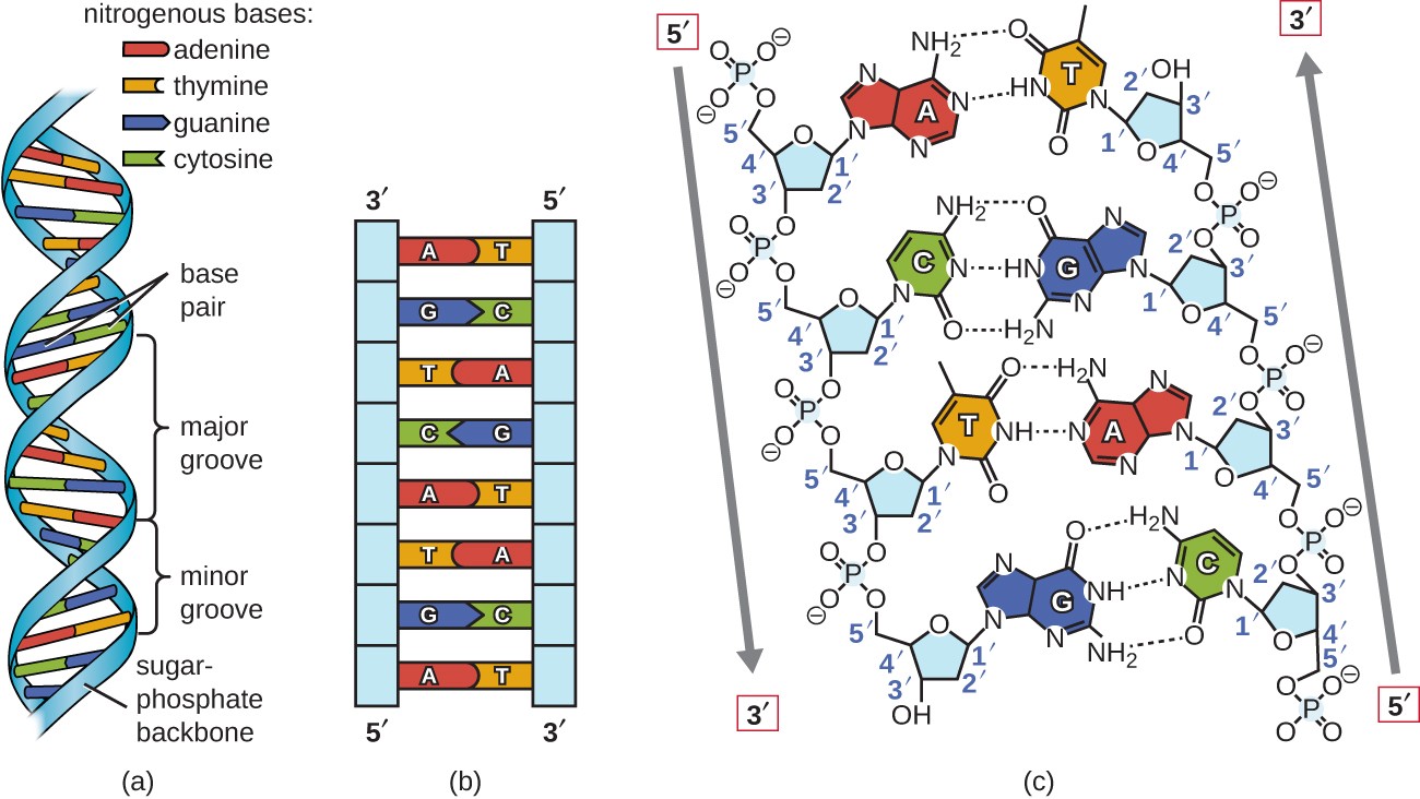
Understanding DNA replication starts with mapping how cellular machinery copies genetic instructions. A clear DNA synthesis diagram breaks down this complex biochemical process into discrete molecular events, showing how enzymes build a complementary strand from a single template.
At the center of this mechanism is the replication fork, where the double helix unwinds and dual strands are copied simultaneously. While the leading strand forms in a continuous sweep, the lagging strand requires short fragments stitched together by specific proteins. Grasping these structural milestones is essential for molecular biology labs, genetic diagnostics, and clinical research.
The Anatomy of the Replication Fork
The replication fork represents the dynamic junction where parental DNA strands separate and new synthesis occurs. This region requires precise coordination among multiple enzymatic actors to maintain structural integrity and speed.
Helicase initiates the process by breaking hydrogen bonds between complementary base pairs. As it moves along the double helix, it separates the strands to expose the genetic code. Single-strand binding proteins coat these exposed segments immediately to prevent them from snapping back together or forming hairpin loops.
Ahead of the advancing fork, torsional stress accumulates as the DNA twists tighter. Topoisomerase relieves this strain by cutting one or both strands, allowing them to swivel and release tension before re-sealing the backbone.
- Helicase: Unzips the parental double helix by disrupting hydrogen bonds.
- Single-Strand Binding Proteins: Stabilize naked template strands against degradation or re-annealing.
- Topoisomerase: Prevents destructive supercoiling by nicking and repairing DNA strands ahead of the fork.
- Primase: Synthesizes short RNA segments to provide starting attachment points.
Leading Versus Lagging Strand Dynamics
Because DNA polymerase only operates in the 5 prime to 3 prime direction, the two template strands demand different handling during replication. This directional constraint creates two distinct pathways at the fork.
The leading strand runs in the correct orientation for the enzyme to follow the opening fork smoothly. Polymerase glides along this template continuously, adding thousands of nucleotides without interruption.
In contrast, the lagging strand runs in the opposite direction. The cell must synthesize this strand backward in short segments known as Okazaki fragments. Each fragment requires a fresh RNA primer laid down by primase before polymerase can extend it toward the previously built segment.
- Leading Strand: Synthesized continuously toward the advancing replication fork.
- Lagging Strand: Synthesized discontinuously away from the fork in short fragments.
- Okazaki Fragments: Small segments of DNA formed on the lagging template strand.
Enzymatic Roles in Chain Elongation
Polymerase acts as the primary catalyst for building the new DNA strand, but it relies heavily on helper proteins to stay on track. Without accessory factors, the enzyme would fall off the template after adding only a few bases.
Sliding clamps form ring-shaped structures that encircle the DNA duplex, tethering the polymerase tightly to the template. A specialized protein complex called the clamp loader uses adenosine triphosphate energy to snap this clamp onto the DNA strand during assembly.
Once replication fragments are complete, RNA primers must be removed. DNA polymerase I excises these temporary RNA pieces and fills the gaps with proper deoxynucleotides. Finally, DNA ligase seals the remaining nicks in the sugar-phosphate backbone, completing the continuous double helix.
- Sliding Clamp: Secures polymerase to the DNA template to enhance speed and efficiency.
- Clamp Loader: Uses chemical energy to position the sliding clamp onto the nucleic acid chain.
- DNA Ligase: Catalyzes the final phosphodiester bond to join adjacent DNA fragments.
Practical Applications in Research and Medicine
Interpreting replication diagrams informs numerous experimental techniques and therapeutic strategies across modern biotechnology.
In diagnostic laboratories, understanding primer placement and polymerase behavior is crucial for designing reliable polymerase chain reaction assays. Medical researchers also target these replication enzymes to develop antiviral medications and chemotherapy drugs. By jamming the synthesis machinery in rapidly dividing cells, treatments can halt viral proliferation or cancer growth.
Synthetic biology pushes these principles further by engineering artificial genomes from scratch. By mastering how enzymes read and copy base sequences, scientists design custom DNA constructs for industrial enzyme production and metabolic engineering.
Frequently Asked Questions
Why does DNA synthesis only occur in one direction?
DNA polymerase requires a free 3 prime hydroxyl group on the growing nucleotide chain to form a chemical bond with an incoming nucleotide's 5 prime phosphate group. The enzyme's active site cannot accept incoming nucleotides from the opposite end, enforcing strict directional synthesis.
What happens if DNA polymerase makes a mistake?
Polymerase features a built-in proofreading function with 3 prime to 5 prime exonuclease activity. If it inserts an incorrect nucleotide, it pauses, clips out the mismatched base, and inserts the correct complementary partner, keeping mutation rates exceptionally low.
How do eukaryotic and prokaryotic replication differ?
Prokaryotes typically feature circular chromosomes with a single origin of replication, managed by simpler enzymatic groups. Eukaryotes possess linear chromosomes with multiple origins, requiring complex histone management and specialized enzymes like telomeres to protect chromosome ends.