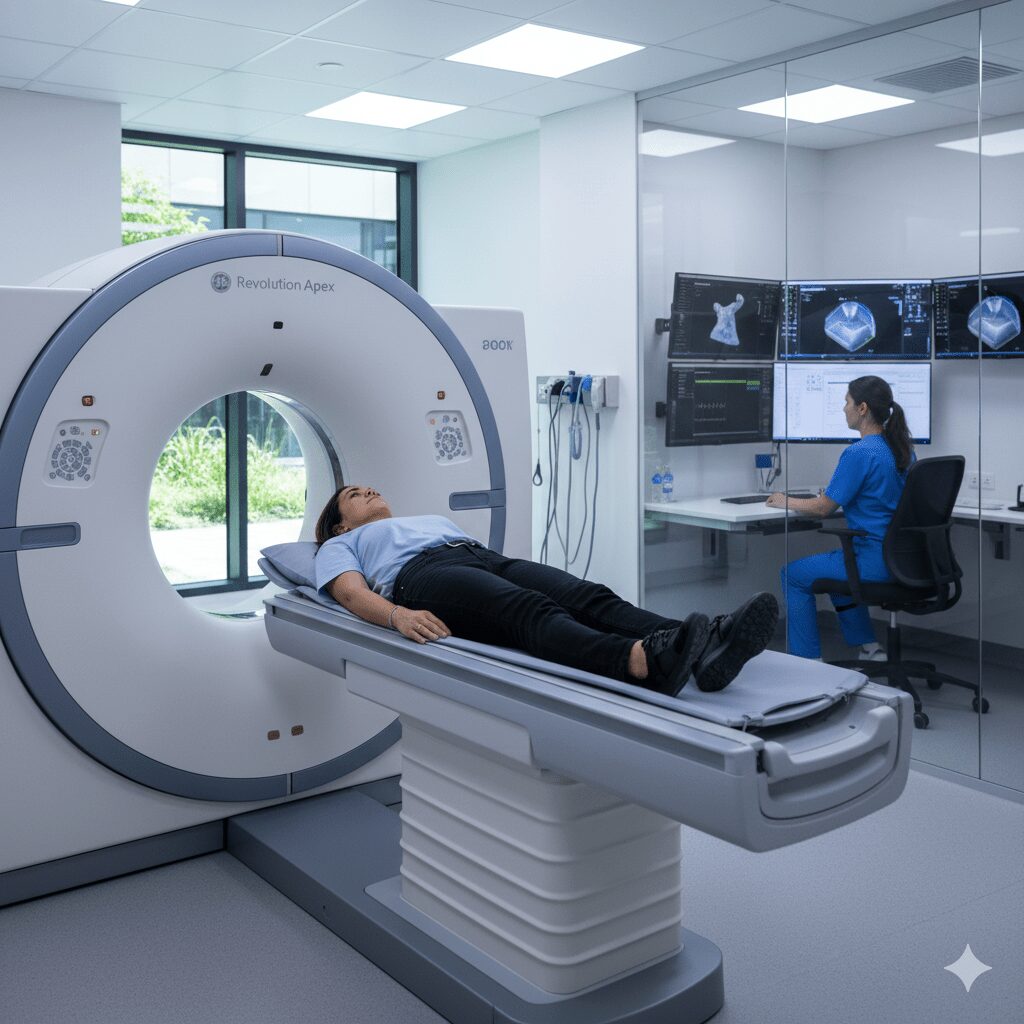
When you study for the Advanced Placement Psychology exam, you quickly run into a wall of clinical jargon. Among the biological bases of behavior, brain imaging techniques often cause the most confusion. A CT scan AP Psychology definition requires you to understand both how the medical technology works and why psychologists use it to study the brain.
At its core, a computed tomography (CT) scan is a computerized X-ray imaging procedure that produces cross-sectional images of the brain. In AP Psychology, you don't need a medical degree in radiology, but you do need to know that a CT scan highlights structural damage, tumors, and gross brain abnormalities. It creates a static picture, showing anatomy rather than activity, which sets it apart from functional scans like fMRIs or PET scans.
What Is a CT Scan in AP Psychology?
A CT scan uses multiple X-ray measurements taken from different angles to produce tomographic images of a specific area of a scanned object. For psychology students, that object is the human brain.
When a patient undergoes a CT scan, an X-ray machine rotates around their head, shooting narrow beams of X-rays through the brain tissue. A computer processes these beams to measure how much radiation is absorbed by different types of tissue. Bone absorbs a lot, appearing white on the final image. Brain tissue absorbs less, appearing in shades of gray. Cerebrospinal fluid-filled ventricles absorb very little, showing up dark.
Structural Imaging vs. Functional Imaging
The single most important distinction an AP Psychology student must master is the difference between structural and functional neuroimaging techniques.
- Structural imaging maps the physical anatomy of the brain. It shows size, shape, and physical damage. CT scans and MRIs fall into this category. They provide a snapshot in time.
- Functional imaging measures brain activity in real time. It shows which brain areas use glucose or oxygen during specific tasks. PET scans and fMRIs fall into this category.
If an exam question asks about locating a physical tumor or a large stroke, the answer involves a structural scan like a CT scan. If the question asks about which brain region lights up when someone experiences fear, the answer requires a functional scan.
How a CT Scan Works
Understanding the mechanics helps anchor your memory for multiple-choice questions. The process relies on differential tissue density.
- Patient Preparation: The patient lies on a motorized table that slides into a doughnut-shaped CT scanner.
- X-Ray Emission: An X-ray source emits a narrow beam through the head to a detector on the opposite side.
- Rotation: The source and detector rotate rapidly around the gantry, taking hundreds of readings per second from different angles.
- Computer Reconstruction: A powerful computer calculates the density of every tiny point in the scanned slice, building a 2D or 3D cross-sectional image.
This non-invasive method allows doctors and researchers to visualize internal structures without surgery. However, it exposes the patient to ionizing radiation, which limits its routine use in purely psychological research settings.
Advantages and Disadvantages in Psychological Research
Every brain imaging technique has distinct trade-offs. Examiners love testing whether you know the strengths and weaknesses of each tool.
Advantages of CT Scans
- Speed: CT scans take only a few minutes, making them ideal for emergency situations where time is critical, such as a suspected stroke or acute head trauma.
- Cost-Effectiveness: They cost significantly less than MRIs or PET scans.
- Accessibility: Most hospitals and major medical clinics have CT scanners, making data collection straightforward for clinical case studies.
- Bone Detail: They provide exceptionally clear images of skull fractures and hard bone structures alongside soft tissue.
Disadvantages for Psychologists
- Radiation Exposure: Because CT scans rely on X-rays, they involve ionizing radiation. Researchers avoid using them in non-medical psychological studies due to these health risks.
- Lower Soft-Tissue Contrast: While a CT scan shows major structural damage, it lacks the fine-grained detail of an MRI when distinguishing between subtle differences in soft brain tissue.
- No Functional Data: A CT scan tells you nothing about brain activity or neurotransmitter function. It's a photograph, not a video.
Common Exam Scenarios and Misconceptions
AP Psychology questions often present a clinical vignette and ask you to select the best imaging tool. Recognizing key trigger words helps you choose correctly.
If a prompt mentions an emergency room patient who fell and might have a skull fracture or bleeding in the brain, the correct answer is almost always a CT scan. If the prompt describes mapping active neural pathways during a memory test, rule out the CT scan immediately and look for an fMRI or PET scan.
A frequent misconception is that CT scans can show psychological disorders like schizophrenia in real time. While research has shown structural brain differences in people with schizophrenia—such as enlarged ventricles—a CT scan only reveals the physical size of those ventricles. It doesn't explain the psychological experience or thought patterns behind the disorder. For more details on psychological assessment standards, you can review guidelines from the American Psychological Association.
Frequently Asked Questions
Does a CT scan show brain function?
No. A CT scan is strictly a structural imaging technique. It reveals the physical anatomy of the brain, such as tissue density, ventricles, and lesions, but it cannot measure neural activity or blood flow changes over time.
Why would a psychologist choose a CT scan over an MRI?
While MRIs provide higher-resolution images of soft tissue without radiation, CT scans are faster, less expensive, and much better at visualizing acute trauma like skull fractures or bleeding.
Are CT scans safe for regular psychological research?
No. Because CT scans use ionizing radiation, institutional review boards and researchers avoid using them for non-diagnostic psychological studies where functional imaging or non-invasive tools like EEG are safer alternatives.
How do CT scans differ from PET scans?
CT scans use X-rays to show brain structure. PET scans use a radioactive tracer substance to measure metabolic activity and glucose consumption, showing which parts of the brain are active.


